Zinc »
PDB 12ca-1add »
1a1r »
Zinc in PDB 1a1r: Hcv NS3 Protease Domain:NS4A Peptide Complex
Protein crystallography data
The structure of Hcv NS3 Protease Domain:NS4A Peptide Complex, PDB code: 1a1r
was solved by
J.L.Kim,
K.A.Morgenstern,
C.Lin,
T.Fox,
M.D.Dwyer,
J.A.Landro,
S.P.Chambers,
W.Markland,
C.A.Lepre,
E.T.O'malley,
S.L.Harbeson,
C.M.Rice,
M.A.Murcko,
P.R.Caron,
J.A.Thomson,
with X-Ray Crystallography technique. A brief refinement statistics is given in the table below:
| Resolution Low / High (Å) | 6.00 / 2.50 |
| Space group | H 3 2 |
| Cell size a, b, c (Å), α, β, γ (°) | 225.000, 225.000, 75.450, 90.00, 90.00, 120.00 |
| R / Rfree (%) | 21.6 / 26.1 |
Zinc Binding Sites:
The binding sites of Zinc atom in the Hcv NS3 Protease Domain:NS4A Peptide Complex
(pdb code 1a1r). This binding sites where shown within
5.0 Angstroms radius around Zinc atom.
In total 2 binding sites of Zinc where determined in the Hcv NS3 Protease Domain:NS4A Peptide Complex, PDB code: 1a1r:
Jump to Zinc binding site number: 1; 2;
In total 2 binding sites of Zinc where determined in the Hcv NS3 Protease Domain:NS4A Peptide Complex, PDB code: 1a1r:
Jump to Zinc binding site number: 1; 2;
Zinc binding site 1 out of 2 in 1a1r
Go back to
Zinc binding site 1 out
of 2 in the Hcv NS3 Protease Domain:NS4A Peptide Complex
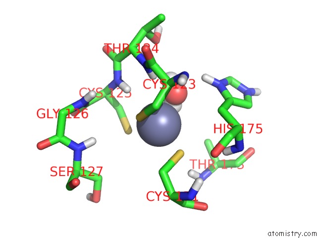
Mono view
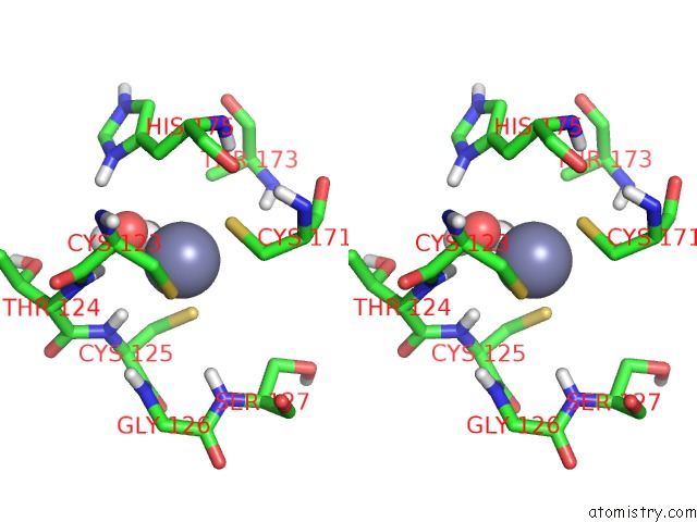
Stereo pair view
Mono view
Stereo pair view
A full contact list of Zinc with other atoms in the Zn binding
site number 1 of Hcv NS3 Protease Domain:NS4A Peptide Complex within 5.0Å range:
|
Zinc binding site 2 out of 2 in 1a1r
Go back to
Zinc binding site 2 out
of 2 in the Hcv NS3 Protease Domain:NS4A Peptide Complex
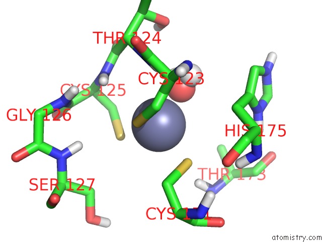
Mono view
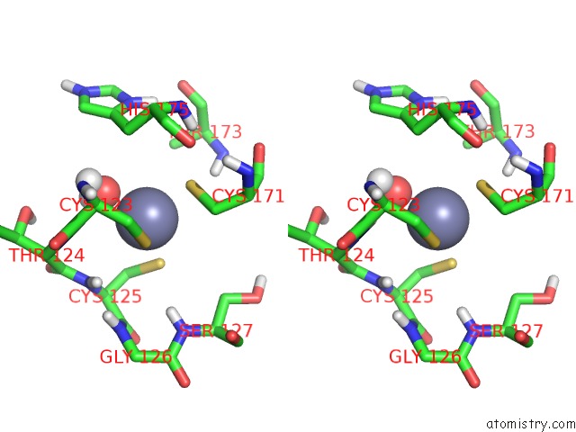
Stereo pair view
Mono view
Stereo pair view
A full contact list of Zinc with other atoms in the Zn binding
site number 2 of Hcv NS3 Protease Domain:NS4A Peptide Complex within 5.0Å range:
|
Reference:
J.L.Kim,
K.A.Morgenstern,
C.Lin,
T.Fox,
M.D.Dwyer,
J.A.Landro,
S.P.Chambers,
W.Markland,
C.A.Lepre,
E.T.O'malley,
S.L.Harbeson,
C.M.Rice,
M.A.Murcko,
P.R.Caron,
J.A.Thomson.
Crystal Structure of the Hepatitis C Virus NS3 Protease Domain Complexed with A Synthetic NS4A Cofactor Peptide. Cell(Cambridge,Mass.) V. 87 343 1996.
ISSN: ISSN 0092-8674
PubMed: 8861917
DOI: 10.1016/S0092-8674(00)81351-3
Page generated: Sat Oct 12 21:44:54 2024
ISSN: ISSN 0092-8674
PubMed: 8861917
DOI: 10.1016/S0092-8674(00)81351-3
Last articles
Zn in 9MJ5Zn in 9HNW
Zn in 9G0L
Zn in 9FNE
Zn in 9DZN
Zn in 9E0I
Zn in 9D32
Zn in 9DAK
Zn in 8ZXC
Zn in 8ZUF