Zinc »
PDB 9ntg-9ueq »
9u42 »
Zinc in PDB 9u42: Crystal Structure of Homogentisate 1,2-Dioxygenase From Acinetobacter in Complex with Zn Ion
Protein crystallography data
The structure of Crystal Structure of Homogentisate 1,2-Dioxygenase From Acinetobacter in Complex with Zn Ion, PDB code: 9u42
was solved by
P.-W.Seo,
S.-A.Hwangbo,
S.-Y.Park,
with X-Ray Crystallography technique. A brief refinement statistics is given in the table below:
| Resolution Low / High (Å) | 24.80 / 1.55 |
| Space group | C 2 2 21 |
| Cell size a, b, c (Å), α, β, γ (°) | 53.058, 121.25, 54.363, 90, 90, 90 |
| R / Rfree (%) | 16.9 / 19.1 |
Zinc Binding Sites:
The binding sites of Zinc atom in the Crystal Structure of Homogentisate 1,2-Dioxygenase From Acinetobacter in Complex with Zn Ion
(pdb code 9u42). This binding sites where shown within
5.0 Angstroms radius around Zinc atom.
In total only one binding site of Zinc was determined in the Crystal Structure of Homogentisate 1,2-Dioxygenase From Acinetobacter in Complex with Zn Ion, PDB code: 9u42:
In total only one binding site of Zinc was determined in the Crystal Structure of Homogentisate 1,2-Dioxygenase From Acinetobacter in Complex with Zn Ion, PDB code: 9u42:
Zinc binding site 1 out of 1 in 9u42
Go back to
Zinc binding site 1 out
of 1 in the Crystal Structure of Homogentisate 1,2-Dioxygenase From Acinetobacter in Complex with Zn Ion
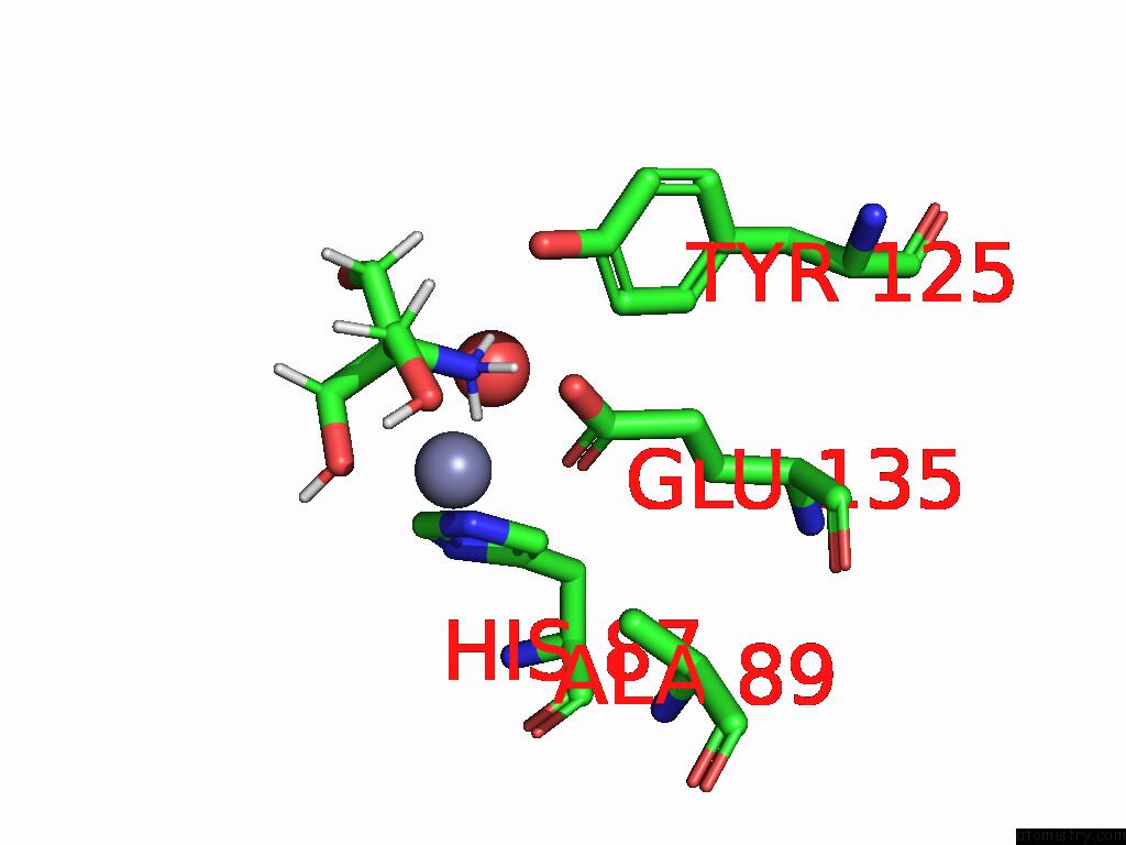
Mono view
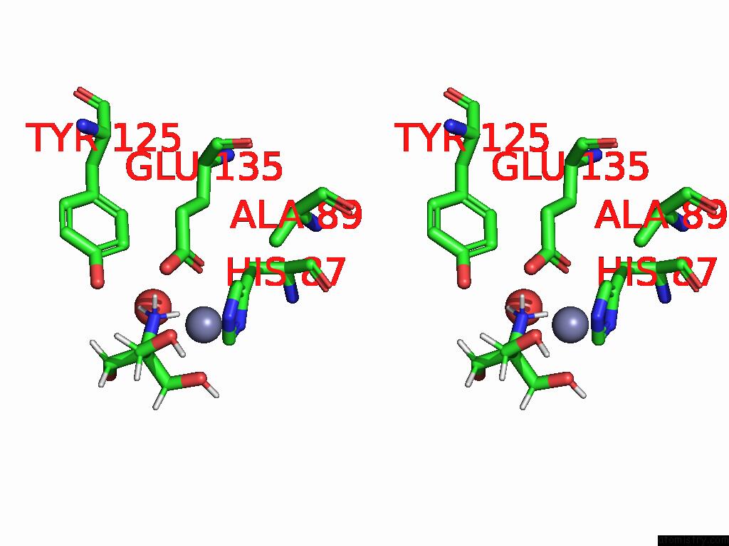
Stereo pair view
Mono view
Stereo pair view
A full contact list of Zinc with other atoms in the Zn binding
site number 1 of Crystal Structure of Homogentisate 1,2-Dioxygenase From Acinetobacter in Complex with Zn Ion within 5.0Å range:
|
Reference:
P.W.Seo,
S.A.Hwangbo,
J.S.Kim,
S.Y.Park.
Structural Mimicry Without Glyoxalase I Functional Convergence: A Homogentisate 1,2-Dioxygenase From Acinetobacter. Proteins 2025.
ISSN: ESSN 1097-0134
PubMed: 40650421
DOI: 10.1002/PROT.70020
Page generated: Fri Aug 22 18:57:42 2025
ISSN: ESSN 1097-0134
PubMed: 40650421
DOI: 10.1002/PROT.70020
Last articles
Ca in 9U8GCa in 9UD8
Ca in 9R0Q
Ca in 9QDT
Ca in 9O4Q
Ca in 9O4O
Ca in 9O4N
Ca in 9O4P
Ca in 9K6M
Ca in 9IUJ