Zinc »
PDB 9ntg-9ueq »
9qi5 »
Zinc in PDB 9qi5: Crystal Structure of I105R Mutant of Blac From Mycobacterium Tuberculosis
Enzymatic activity of Crystal Structure of I105R Mutant of Blac From Mycobacterium Tuberculosis
All present enzymatic activity of Crystal Structure of I105R Mutant of Blac From Mycobacterium Tuberculosis:
3.5.2.6;
3.5.2.6;
Protein crystallography data
The structure of Crystal Structure of I105R Mutant of Blac From Mycobacterium Tuberculosis, PDB code: 9qi5
was solved by
A.Chikunova,
M.Radojkovic,
M.Ubbink,
with X-Ray Crystallography technique. A brief refinement statistics is given in the table below:
| Resolution Low / High (Å) | 44.62 / 1.80 |
| Space group | P 21 21 21 |
| Cell size a, b, c (Å), α, β, γ (°) | 39.161, 40.975, 267.495, 90, 90, 90 |
| R / Rfree (%) | 17.4 / 21.1 |
Zinc Binding Sites:
The binding sites of Zinc atom in the Crystal Structure of I105R Mutant of Blac From Mycobacterium Tuberculosis
(pdb code 9qi5). This binding sites where shown within
5.0 Angstroms radius around Zinc atom.
In total only one binding site of Zinc was determined in the Crystal Structure of I105R Mutant of Blac From Mycobacterium Tuberculosis, PDB code: 9qi5:
In total only one binding site of Zinc was determined in the Crystal Structure of I105R Mutant of Blac From Mycobacterium Tuberculosis, PDB code: 9qi5:
Zinc binding site 1 out of 1 in 9qi5
Go back to
Zinc binding site 1 out
of 1 in the Crystal Structure of I105R Mutant of Blac From Mycobacterium Tuberculosis
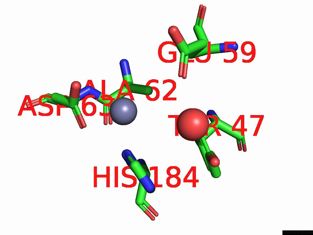
Mono view
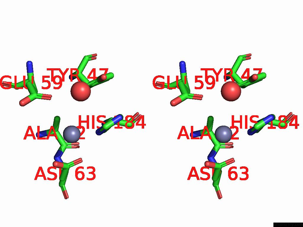
Stereo pair view
Mono view
Stereo pair view
A full contact list of Zinc with other atoms in the Zn binding
site number 1 of Crystal Structure of I105R Mutant of Blac From Mycobacterium Tuberculosis within 5.0Å range:
|
Reference:
M.Radojkovic,
S.F.Koene,
A.Chikunova,
M.Timmer,
S.V.Natarajan,
M.Ubbink.
High-Resolution Activity and Inhibitor Profiling of Residue 105 in Class A Beta-Lactamases To Be Published.
Page generated: Fri Aug 22 18:56:14 2025
Last articles
Ca in 9K6MCa in 9IUJ
Ca in 9JD0
Ca in 9JD1
Ca in 9JCX
Ca in 9I6C
Ca in 9GTB
Ca in 9GSH
Ca in 9GSI
Ca in 9GRG