Zinc »
PDB 9ntg-9ueq »
9qdh »
Zinc in PDB 9qdh: Crystal Structure of Iga Protease (323-878) From Thomasclavelia Ramosa
Protein crystallography data
The structure of Crystal Structure of Iga Protease (323-878) From Thomasclavelia Ramosa, PDB code: 9qdh
was solved by
M.A.Marquez-Monino,
A.Martinez Gascuena,
M.Aguillo-Urarte,
A.Manzanares-Gomez,
B.Trastoy,
with X-Ray Crystallography technique. A brief refinement statistics is given in the table below:
| Resolution Low / High (Å) | 55.77 / 2.00 |
| Space group | P 21 21 21 |
| Cell size a, b, c (Å), α, β, γ (°) | 70.739, 85.198, 90.666, 90, 90, 90 |
| R / Rfree (%) | 19.4 / 24.5 |
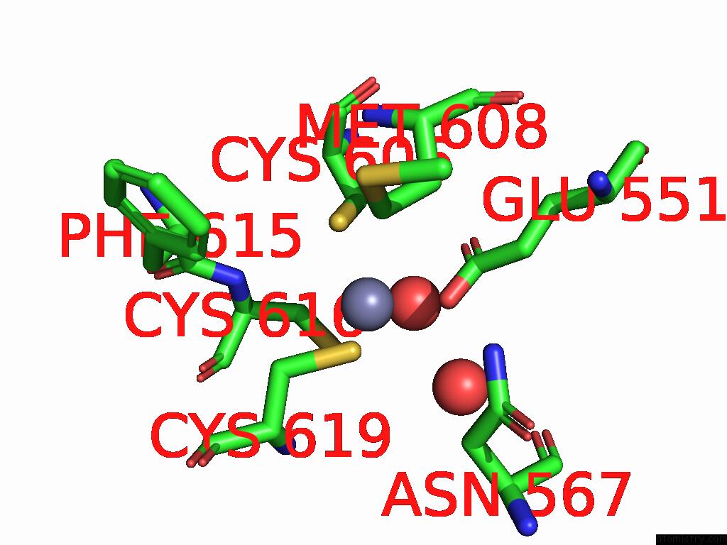
Zinc Binding Sites:
The binding sites of Zinc atom in the Crystal Structure of Iga Protease (323-878) From Thomasclavelia Ramosa
(pdb code 9qdh). This binding sites where shown within
5.0 Angstroms radius around Zinc atom.
In total only one binding site of Zinc was determined in the Crystal Structure of Iga Protease (323-878) From Thomasclavelia Ramosa, PDB code: 9qdh:
In total only one binding site of Zinc was determined in the Crystal Structure of Iga Protease (323-878) From Thomasclavelia Ramosa, PDB code: 9qdh:
Zinc binding site 1 out of 1 in 9qdh
Go back to
Zinc binding site 1 out
of 1 in the Crystal Structure of Iga Protease (323-878) From Thomasclavelia Ramosa
Mono view
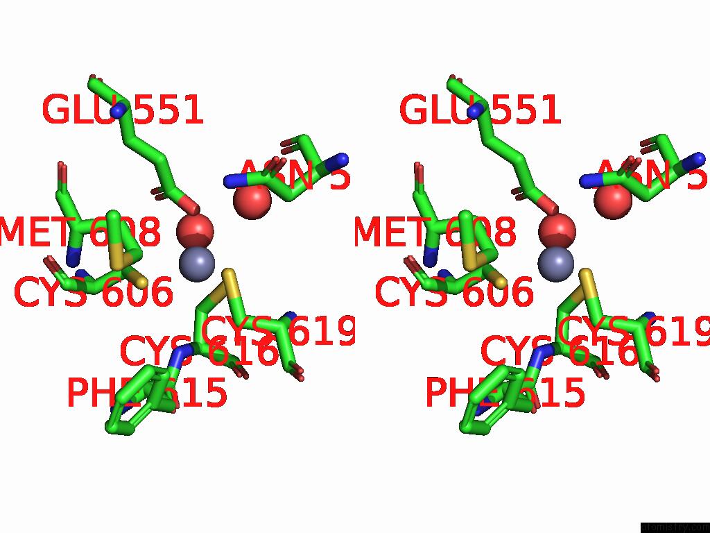
Stereo pair view
Mono view
Stereo pair view
A full contact list of Zinc with other atoms in the Zn binding
site number 1 of Crystal Structure of Iga Protease (323-878) From Thomasclavelia Ramosa within 5.0Å range:
|
Reference:
M.A.Marquez-Monino,
A.Martinez Gascuena,
T.Azzam,
A.Pesson,
A.Manzanares-Gomez,
M.Aguillo-Urarte,
T.T.Brown,
A.Montero,
R.Lood,
A.Naegali,
J.J.Du,
S.R.Connell,
D.E.Sastre,
E.J.Sundberg,
B.Trastoy.
Structural Insights Into Iga Recognition By M64 Peptidases To Be Published.
Page generated: Fri Aug 22 18:55:42 2025
Last articles
Ca in 9VAKCa in 9O9V
Ca in 9U8G
Ca in 9UD8
Ca in 9R0Q
Ca in 9QDT
Ca in 9O4Q
Ca in 9O4O
Ca in 9O4N
Ca in 9O4P