Zinc »
PDB 7kmm-7l3l »
7ky2 »
Zinc in PDB 7ky2: Botulism Neurooxin Light Chain A App Form
Protein crystallography data
The structure of Botulism Neurooxin Light Chain A App Form, PDB code: 7ky2
was solved by
M.E.Ortega,
N.T.Salazameda,
with X-Ray Crystallography technique. A brief refinement statistics is given in the table below:
| Resolution Low / High (Å) | 49.01 / 2.78 |
| Space group | P 1 21 1 |
| Cell size a, b, c (Å), α, β, γ (°) | 39.029, 56.985, 192.136, 90, 90.26, 90 |
| R / Rfree (%) | 12.8 / 17 |
Zinc Binding Sites:
The binding sites of Zinc atom in the Botulism Neurooxin Light Chain A App Form
(pdb code 7ky2). This binding sites where shown within
5.0 Angstroms radius around Zinc atom.
In total 2 binding sites of Zinc where determined in the Botulism Neurooxin Light Chain A App Form, PDB code: 7ky2:
Jump to Zinc binding site number: 1; 2;
In total 2 binding sites of Zinc where determined in the Botulism Neurooxin Light Chain A App Form, PDB code: 7ky2:
Jump to Zinc binding site number: 1; 2;
Zinc binding site 1 out of 2 in 7ky2
Go back to
Zinc binding site 1 out
of 2 in the Botulism Neurooxin Light Chain A App Form
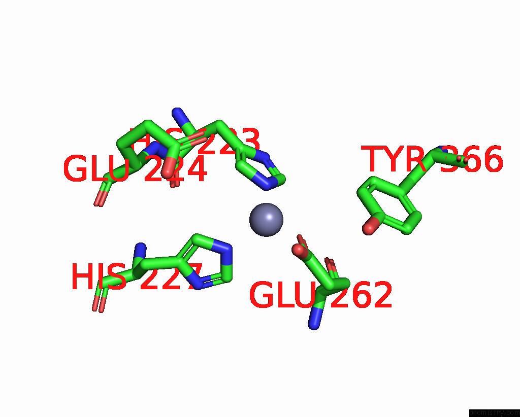
Mono view
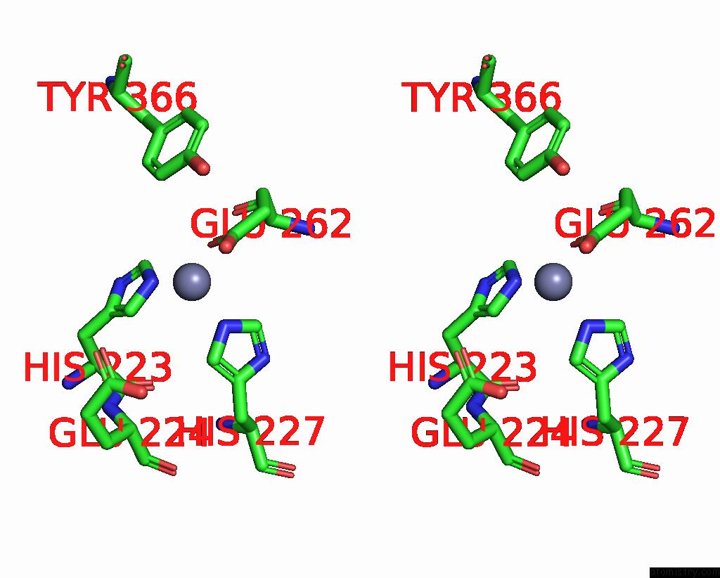
Stereo pair view
Mono view
Stereo pair view
A full contact list of Zinc with other atoms in the Zn binding
site number 1 of Botulism Neurooxin Light Chain A App Form within 5.0Å range:
|
Zinc binding site 2 out of 2 in 7ky2
Go back to
Zinc binding site 2 out
of 2 in the Botulism Neurooxin Light Chain A App Form

Mono view
Stereo pair view
Mono view
Stereo pair view
A full contact list of Zinc with other atoms in the Zn binding
site number 2 of Botulism Neurooxin Light Chain A App Form within 5.0Å range:
|
Reference:
M.Amezcua,
R.S.Cruz,
A.Ku,
W.Moran,
M.E.Ortega,
N.T.Salzameda.
Discovery of Dipeptides As Potent Botulinum Neurotoxin A Light-Chain Inhibitors. Acs Med.Chem.Lett. V. 12 295 2021.
ISSN: ISSN 1948-5875
PubMed: 33603978
DOI: 10.1021/ACSMEDCHEMLETT.0C00674
Page generated: Fri Aug 22 01:44:33 2025
ISSN: ISSN 1948-5875
PubMed: 33603978
DOI: 10.1021/ACSMEDCHEMLETT.0C00674
Last articles
Mn in 9LJUMn in 9LJW
Mn in 9LJS
Mn in 9LJR
Mn in 9LJT
Mn in 9LJV
Mg in 9UA2
Mg in 9R96
Mg in 9VM1
Mg in 9P01