Zinc »
PDB 7hl3-7jnz »
7hoc »
Zinc in PDB 7hoc: Group Deposition For Crystallographic Fragment Screening of Coxsackievirus A16 (G-10) 2A Protease -- Crystal Structure of Coxsackievirus A16 (G-10) 2A Protease in Complex with Z802821712 (A71EV2A-X0437)
Enzymatic activity of Group Deposition For Crystallographic Fragment Screening of Coxsackievirus A16 (G-10) 2A Protease -- Crystal Structure of Coxsackievirus A16 (G-10) 2A Protease in Complex with Z802821712 (A71EV2A-X0437)
All present enzymatic activity of Group Deposition For Crystallographic Fragment Screening of Coxsackievirus A16 (G-10) 2A Protease -- Crystal Structure of Coxsackievirus A16 (G-10) 2A Protease in Complex with Z802821712 (A71EV2A-X0437):
3.4.22.29;
3.4.22.29;
Protein crystallography data
The structure of Group Deposition For Crystallographic Fragment Screening of Coxsackievirus A16 (G-10) 2A Protease -- Crystal Structure of Coxsackievirus A16 (G-10) 2A Protease in Complex with Z802821712 (A71EV2A-X0437), PDB code: 7hoc
was solved by
R.M.Lithgo,
M.Fairhead,
L.Koekemoer,
B.H.Balcomb,
E.Capkin,
A.V.Chandran,
M.Golding,
A.S.Godoy,
J.C.Aschenbrenner,
P.G.Marples,
X.Ni,
W.Thompson,
C.W.E.Tomlinson,
C.Wild,
M.Winokan,
M.-A.E.Xavier,
D.Fearon,
F.Von Delft,
with X-Ray Crystallography technique. A brief refinement statistics is given in the table below:
| Resolution Low / High (Å) | 47.21 / 1.40 |
| Space group | C 1 2 1 |
| Cell size a, b, c (Å), α, β, γ (°) | 86.27, 56.4, 64.53, 90, 94.4, 90 |
| R / Rfree (%) | 19.7 / 23.2 |
Zinc Binding Sites:
The binding sites of Zinc atom in the Group Deposition For Crystallographic Fragment Screening of Coxsackievirus A16 (G-10) 2A Protease -- Crystal Structure of Coxsackievirus A16 (G-10) 2A Protease in Complex with Z802821712 (A71EV2A-X0437)
(pdb code 7hoc). This binding sites where shown within
5.0 Angstroms radius around Zinc atom.
In total 2 binding sites of Zinc where determined in the Group Deposition For Crystallographic Fragment Screening of Coxsackievirus A16 (G-10) 2A Protease -- Crystal Structure of Coxsackievirus A16 (G-10) 2A Protease in Complex with Z802821712 (A71EV2A-X0437), PDB code: 7hoc:
Jump to Zinc binding site number: 1; 2;
Zinc binding site 1 out of 2 in 7hoc
Go back to
Zinc binding site 1 out
of 2 in the Group Deposition For Crystallographic Fragment Screening of Coxsackievirus A16 (G-10) 2A Protease -- Crystal Structure of Coxsackievirus A16 (G-10) 2A Protease in Complex with Z802821712 (A71EV2A-X0437)
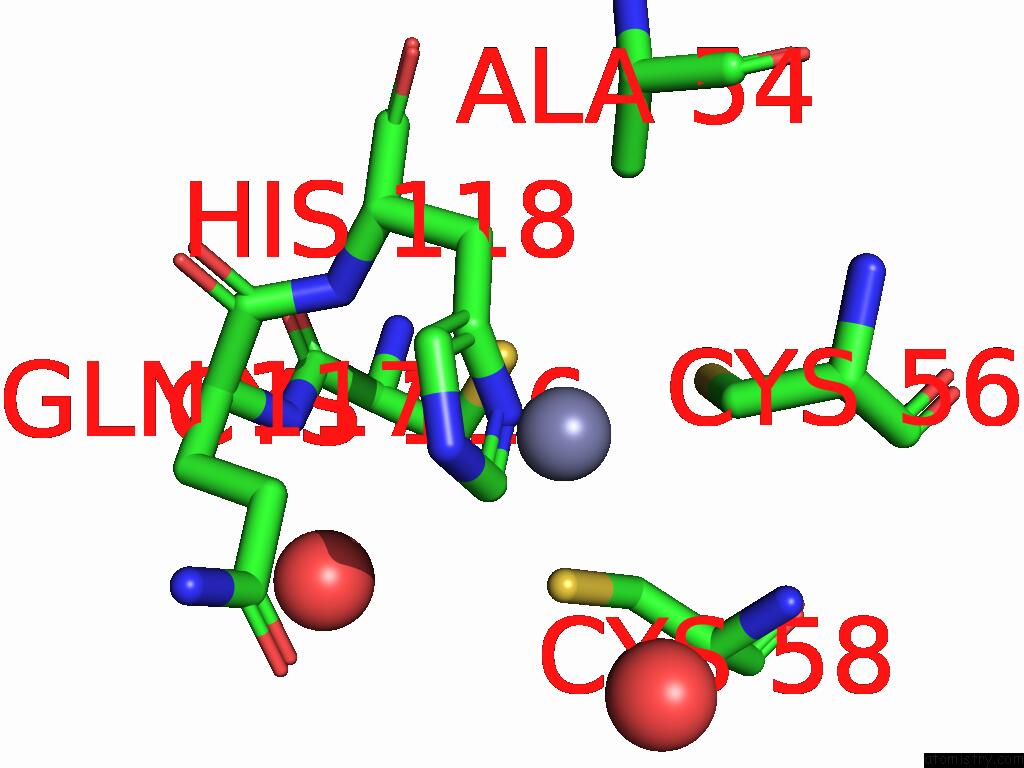
Mono view
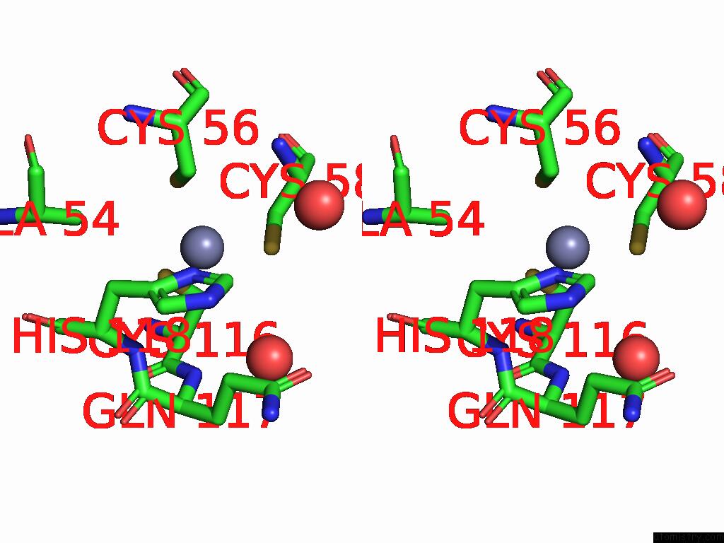
Stereo pair view
Mono view
Stereo pair view
A full contact list of Zinc with other atoms in the Zn binding
site number 1 of Group Deposition For Crystallographic Fragment Screening of Coxsackievirus A16 (G-10) 2A Protease -- Crystal Structure of Coxsackievirus A16 (G-10) 2A Protease in Complex with Z802821712 (A71EV2A-X0437) within 5.0Å range:
|
Zinc binding site 2 out of 2 in 7hoc
Go back to
Zinc binding site 2 out
of 2 in the Group Deposition For Crystallographic Fragment Screening of Coxsackievirus A16 (G-10) 2A Protease -- Crystal Structure of Coxsackievirus A16 (G-10) 2A Protease in Complex with Z802821712 (A71EV2A-X0437)
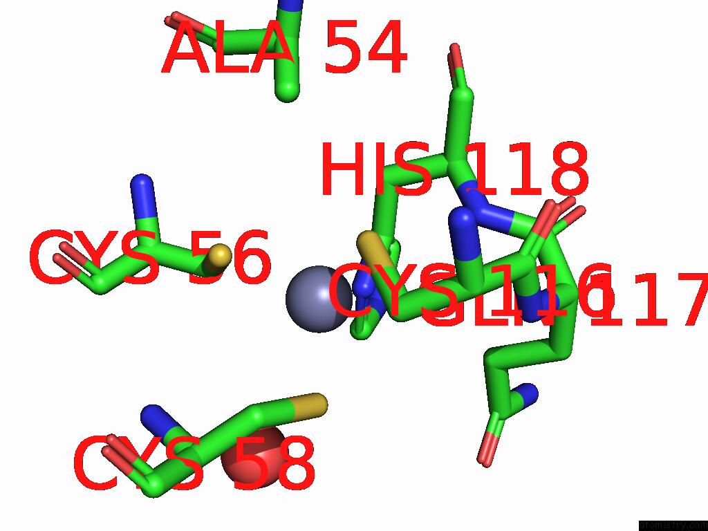
Mono view
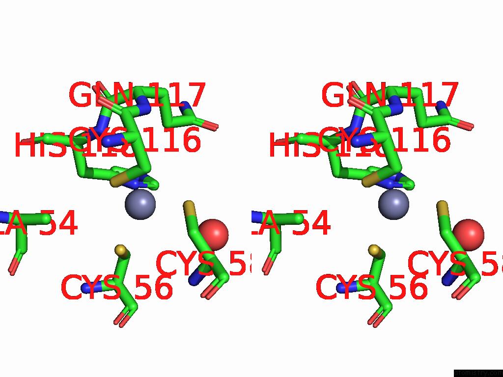
Stereo pair view
Mono view
Stereo pair view
A full contact list of Zinc with other atoms in the Zn binding
site number 2 of Group Deposition For Crystallographic Fragment Screening of Coxsackievirus A16 (G-10) 2A Protease -- Crystal Structure of Coxsackievirus A16 (G-10) 2A Protease in Complex with Z802821712 (A71EV2A-X0437) within 5.0Å range:
|
Reference:
R.M.Lithgo,
M.Fairhead,
L.Koekemoer,
B.H.Balcomb,
E.Capkin,
A.V.Chandran,
M.Golding,
A.S.Godoy,
J.C.Aschenbrenner,
P.G.Marples,
X.Ni,
W.Thompson,
C.W.E.Tomlinson,
C.Wild,
M.Winokan,
M.-A.E.Xavier,
D.Fearon,
F.Von Delft.
Group Deposition For Crystallographic Fragment Screening of Coxsackievirus A16 (G-10) 2A Protease To Be Published.
Page generated: Sun Feb 9 00:41:24 2025
Last articles
Fe in 2YXOFe in 2YRS
Fe in 2YXC
Fe in 2YNM
Fe in 2YVJ
Fe in 2YP1
Fe in 2YU2
Fe in 2YU1
Fe in 2YQB
Fe in 2YOO