Zinc »
PDB 7ecc-7eqz »
7ef9 »
Zinc in PDB 7ef9: Crystal Structure of Mouse Mutyh in Complex with Dna Containing Ap Site Analogue:8-Oxog (Form II)
Enzymatic activity of Crystal Structure of Mouse Mutyh in Complex with Dna Containing Ap Site Analogue:8-Oxog (Form II)
All present enzymatic activity of Crystal Structure of Mouse Mutyh in Complex with Dna Containing Ap Site Analogue:8-Oxog (Form II):
3.2.2.31;
3.2.2.31;
Protein crystallography data
The structure of Crystal Structure of Mouse Mutyh in Complex with Dna Containing Ap Site Analogue:8-Oxog (Form II), PDB code: 7ef9
was solved by
T.Nakamura,
Y.Nakabeppu,
Y.Yamagata,
with X-Ray Crystallography technique. A brief refinement statistics is given in the table below:
| Resolution Low / High (Å) | 42.53 / 1.97 |
| Space group | I 2 2 2 |
| Cell size a, b, c (Å), α, β, γ (°) | 71.66, 108.496, 158.527, 90, 90, 90 |
| R / Rfree (%) | 17.8 / 19.7 |
Other elements in 7ef9:
The structure of Crystal Structure of Mouse Mutyh in Complex with Dna Containing Ap Site Analogue:8-Oxog (Form II) also contains other interesting chemical elements:
| Iron | (Fe) | 4 atoms |
Zinc Binding Sites:
The binding sites of Zinc atom in the Crystal Structure of Mouse Mutyh in Complex with Dna Containing Ap Site Analogue:8-Oxog (Form II)
(pdb code 7ef9). This binding sites where shown within
5.0 Angstroms radius around Zinc atom.
In total only one binding site of Zinc was determined in the Crystal Structure of Mouse Mutyh in Complex with Dna Containing Ap Site Analogue:8-Oxog (Form II), PDB code: 7ef9:
In total only one binding site of Zinc was determined in the Crystal Structure of Mouse Mutyh in Complex with Dna Containing Ap Site Analogue:8-Oxog (Form II), PDB code: 7ef9:
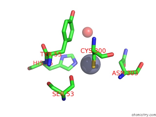
Zinc binding site 1 out of 1 in 7ef9
Go back to
Zinc binding site 1 out
of 1 in the Crystal Structure of Mouse Mutyh in Complex with Dna Containing Ap Site Analogue:8-Oxog (Form II)
Mono view

Stereo pair view
Mono view
Stereo pair view
A full contact list of Zinc with other atoms in the Zn binding
site number 1 of Crystal Structure of Mouse Mutyh in Complex with Dna Containing Ap Site Analogue:8-Oxog (Form II) within 5.0Å range:
|
Reference:
T.Nakamura,
K.Okabe,
S.Hirayama,
M.Chirifu,
S.Ikemizu,
H.Morioka,
Y.Nakabeppu,
Y.Yamagata.
Structure of the Mammalian Adenine Dna Glycosylase Mutyh: Insights Into the Base Excision Repair Pathway and Cancer. Nucleic Acids Res. 2021.
ISSN: ESSN 1362-4962
DOI: 10.1093/NAR/GKAB492
Page generated: Tue Oct 29 19:44:30 2024
ISSN: ESSN 1362-4962
DOI: 10.1093/NAR/GKAB492
Last articles
Mg in 2YEJMg in 2YEF
Mg in 2YE8
Mg in 2YE1
Mg in 2YCH
Mg in 2YCF
Mg in 2YC5
Mg in 2YBD
Mg in 2YC4
Mg in 2YBE