Zinc »
PDB 7del-7dox »
7dgo »
Zinc in PDB 7dgo: The Zn-Bound Dimeric Structure of K79H/G80A/H81A Myoglobin
Protein crystallography data
The structure of The Zn-Bound Dimeric Structure of K79H/G80A/H81A Myoglobin, PDB code: 7dgo
was solved by
S.Nagao,
A.Idomoto,
N.Shibata,
Y.Higuchi,
S.Hirota,
with X-Ray Crystallography technique. A brief refinement statistics is given in the table below:
| Resolution Low / High (Å) | 43.98 / 2.00 |
| Space group | P 21 21 21 |
| Cell size a, b, c (Å), α, β, γ (°) | 52.738, 63.141, 79.466, 90, 90, 90 |
| R / Rfree (%) | 21.3 / 26.5 |
Other elements in 7dgo:
The structure of The Zn-Bound Dimeric Structure of K79H/G80A/H81A Myoglobin also contains other interesting chemical elements:
| Iron | (Fe) | 2 atoms |
Zinc Binding Sites:
The binding sites of Zinc atom in the The Zn-Bound Dimeric Structure of K79H/G80A/H81A Myoglobin
(pdb code 7dgo). This binding sites where shown within
5.0 Angstroms radius around Zinc atom.
In total 5 binding sites of Zinc where determined in the The Zn-Bound Dimeric Structure of K79H/G80A/H81A Myoglobin, PDB code: 7dgo:
Jump to Zinc binding site number: 1; 2; 3; 4; 5;
In total 5 binding sites of Zinc where determined in the The Zn-Bound Dimeric Structure of K79H/G80A/H81A Myoglobin, PDB code: 7dgo:
Jump to Zinc binding site number: 1; 2; 3; 4; 5;
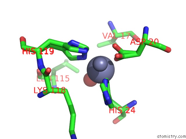
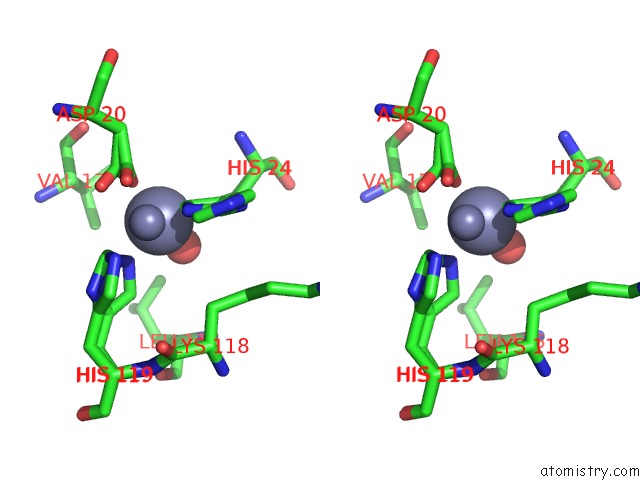
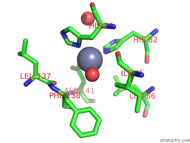
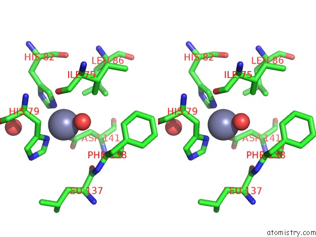
Zinc binding site 1 out of 5 in 7dgo
Go back to
Zinc binding site 1 out
of 5 in the The Zn-Bound Dimeric Structure of K79H/G80A/H81A Myoglobin
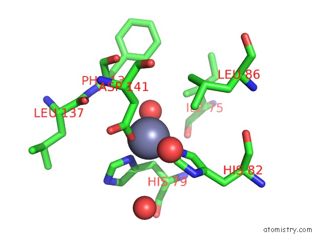
Mono view
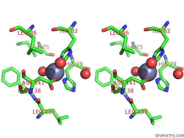
Stereo pair view
Mono view
Stereo pair view
A full contact list of Zinc with other atoms in the Zn binding
site number 1 of The Zn-Bound Dimeric Structure of K79H/G80A/H81A Myoglobin within 5.0Å range:
|
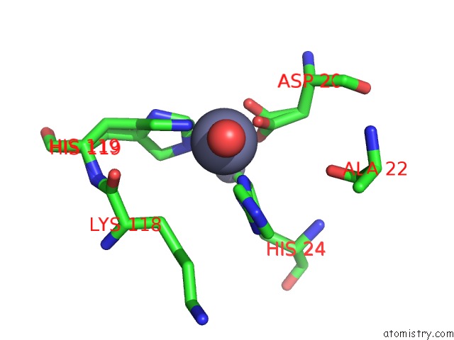
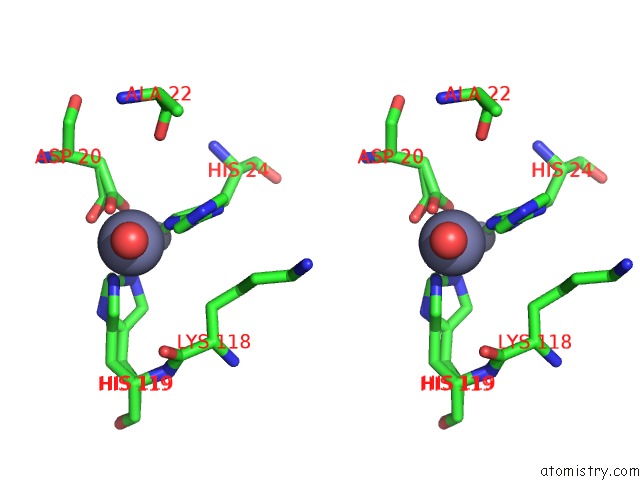
Zinc binding site 2 out of 5 in 7dgo
Go back to
Zinc binding site 2 out
of 5 in the The Zn-Bound Dimeric Structure of K79H/G80A/H81A Myoglobin
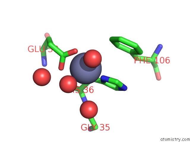
Mono view
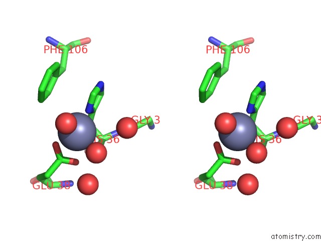
Stereo pair view
Mono view
Stereo pair view
A full contact list of Zinc with other atoms in the Zn binding
site number 2 of The Zn-Bound Dimeric Structure of K79H/G80A/H81A Myoglobin within 5.0Å range:
|
Zinc binding site 3 out of 5 in 7dgo
Go back to
Zinc binding site 3 out
of 5 in the The Zn-Bound Dimeric Structure of K79H/G80A/H81A Myoglobin
Mono view
Stereo pair view
Mono view
Stereo pair view
A full contact list of Zinc with other atoms in the Zn binding
site number 3 of The Zn-Bound Dimeric Structure of K79H/G80A/H81A Myoglobin within 5.0Å range:
|
Zinc binding site 4 out of 5 in 7dgo
Go back to
Zinc binding site 4 out
of 5 in the The Zn-Bound Dimeric Structure of K79H/G80A/H81A Myoglobin
Mono view
Stereo pair view
Mono view
Stereo pair view
A full contact list of Zinc with other atoms in the Zn binding
site number 4 of The Zn-Bound Dimeric Structure of K79H/G80A/H81A Myoglobin within 5.0Å range:
|
Zinc binding site 5 out of 5 in 7dgo
Go back to
Zinc binding site 5 out
of 5 in the The Zn-Bound Dimeric Structure of K79H/G80A/H81A Myoglobin
Mono view
Stereo pair view
Mono view
Stereo pair view
A full contact list of Zinc with other atoms in the Zn binding
site number 5 of The Zn-Bound Dimeric Structure of K79H/G80A/H81A Myoglobin within 5.0Å range:
|
Reference:
S.Nagao,
A.Idomoto,
N.Shibata,
Y.Higuchi,
S.Hirota.
Rational Design of Metal-Binding Sites in Domain-Swapped Myoglobin Dimers. J.Inorg.Biochem. 2021.
ISSN: ISSN 0162-0134
DOI: 10.1016/J.JINORGBIO.2021.111374
Page generated: Tue Oct 29 18:57:18 2024
ISSN: ISSN 0162-0134
DOI: 10.1016/J.JINORGBIO.2021.111374
Last articles
Mg in 5LB5Mg in 5LB3
Mg in 5LAQ
Mg in 5LA6
Mg in 5LA3
Mg in 5L9H
Mg in 5L8F
Mg in 5L9N
Mg in 5L8Y
Mg in 5L6C