Zinc »
PDB 7d59-7dem »
7d8e »
Zinc in PDB 7d8e: Crystal Structure of Double Mutant Y115E Y117E Human Secretory Glutaminyl Cyclase in Complex with Lsb-09
Enzymatic activity of Crystal Structure of Double Mutant Y115E Y117E Human Secretory Glutaminyl Cyclase in Complex with Lsb-09
All present enzymatic activity of Crystal Structure of Double Mutant Y115E Y117E Human Secretory Glutaminyl Cyclase in Complex with Lsb-09:
2.3.2.5;
2.3.2.5;
Protein crystallography data
The structure of Crystal Structure of Double Mutant Y115E Y117E Human Secretory Glutaminyl Cyclase in Complex with Lsb-09, PDB code: 7d8e
was solved by
K.V.Dileep,
K.Ihara,
N.Sakai,
M.Shirozu,
K.Y.J.Zhang,
with X-Ray Crystallography technique. A brief refinement statistics is given in the table below:
| Resolution Low / High (Å) | 46.85 / 2.00 |
| Space group | C 1 2 1 |
| Cell size a, b, c (Å), α, β, γ (°) | 86.181, 149.303, 94.539, 90, 97.64, 90 |
| R / Rfree (%) | 20.9 / 25.2 |
Zinc Binding Sites:
The binding sites of Zinc atom in the Crystal Structure of Double Mutant Y115E Y117E Human Secretory Glutaminyl Cyclase in Complex with Lsb-09
(pdb code 7d8e). This binding sites where shown within
5.0 Angstroms radius around Zinc atom.
In total 3 binding sites of Zinc where determined in the Crystal Structure of Double Mutant Y115E Y117E Human Secretory Glutaminyl Cyclase in Complex with Lsb-09, PDB code: 7d8e:
Jump to Zinc binding site number: 1; 2; 3;
In total 3 binding sites of Zinc where determined in the Crystal Structure of Double Mutant Y115E Y117E Human Secretory Glutaminyl Cyclase in Complex with Lsb-09, PDB code: 7d8e:
Jump to Zinc binding site number: 1; 2; 3;
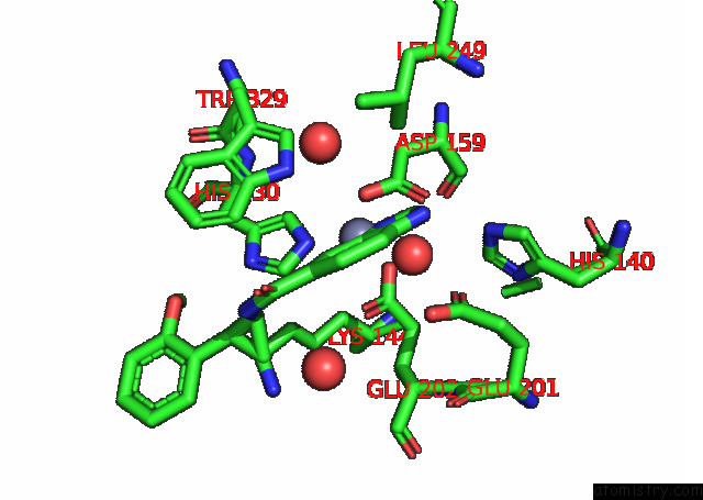
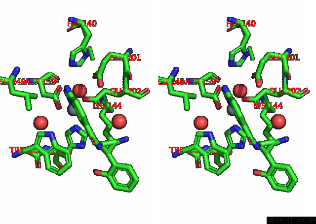
Zinc binding site 1 out of 3 in 7d8e
Go back to
Zinc binding site 1 out
of 3 in the Crystal Structure of Double Mutant Y115E Y117E Human Secretory Glutaminyl Cyclase in Complex with Lsb-09
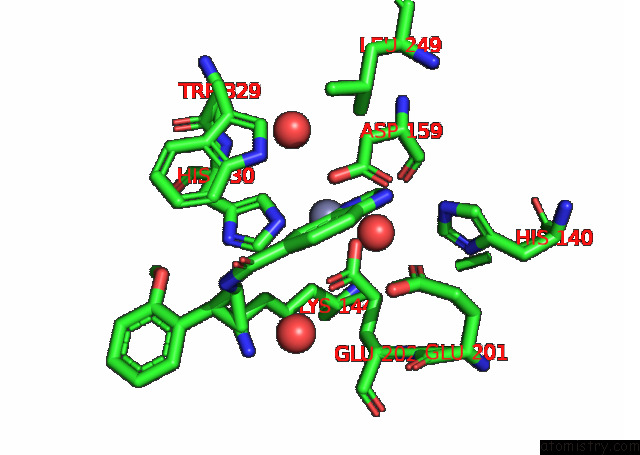
Mono view
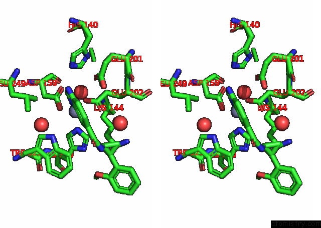
Stereo pair view
Mono view
Stereo pair view
A full contact list of Zinc with other atoms in the Zn binding
site number 1 of Crystal Structure of Double Mutant Y115E Y117E Human Secretory Glutaminyl Cyclase in Complex with Lsb-09 within 5.0Å range:
|
Zinc binding site 2 out of 3 in 7d8e
Go back to
Zinc binding site 2 out
of 3 in the Crystal Structure of Double Mutant Y115E Y117E Human Secretory Glutaminyl Cyclase in Complex with Lsb-09
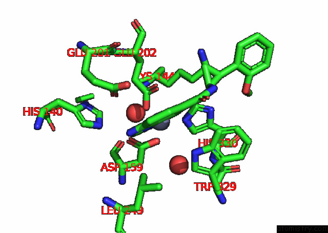
Mono view
Stereo pair view
Mono view
Stereo pair view
A full contact list of Zinc with other atoms in the Zn binding
site number 2 of Crystal Structure of Double Mutant Y115E Y117E Human Secretory Glutaminyl Cyclase in Complex with Lsb-09 within 5.0Å range:
|
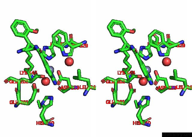
Zinc binding site 3 out of 3 in 7d8e
Go back to
Zinc binding site 3 out
of 3 in the Crystal Structure of Double Mutant Y115E Y117E Human Secretory Glutaminyl Cyclase in Complex with Lsb-09
Mono view
Stereo pair view
Mono view
Stereo pair view
A full contact list of Zinc with other atoms in the Zn binding
site number 3 of Crystal Structure of Double Mutant Y115E Y117E Human Secretory Glutaminyl Cyclase in Complex with Lsb-09 within 5.0Å range:
|
Reference:
K.V.Dileep,
K.Ihara,
N.Sakai,
M.Shirozu,
K.Y.J.Zhang.
Crystal Structure of Double Mutant Y115E Y117E Human Secretory Glutaminyl Cyclase in Complex with Lsb-09 To Be Published.
Page generated: Thu Aug 21 23:25:27 2025
Last articles
Cd in 9J00Cd in 9J01
Ca in 9VAK
Ca in 9O9V
Ca in 9U8G
Ca in 9UD8
Ca in 9R0Q
Ca in 9QDT
Ca in 9O4Q
Ca in 9O4O