Zinc »
PDB 7ci5-7crp »
7cp0 »
Zinc in PDB 7cp0: Crystal Structure of Double Mutant Y115E Y117E Human Secretory Glutaminyl Cyclase
Enzymatic activity of Crystal Structure of Double Mutant Y115E Y117E Human Secretory Glutaminyl Cyclase
All present enzymatic activity of Crystal Structure of Double Mutant Y115E Y117E Human Secretory Glutaminyl Cyclase:
2.3.2.5;
2.3.2.5;
Protein crystallography data
The structure of Crystal Structure of Double Mutant Y115E Y117E Human Secretory Glutaminyl Cyclase, PDB code: 7cp0
was solved by
K.V.Dileep,
K.Ihara,
N.Sakai,
M.Shirozu,
K.Y.J.Zhang,
with X-Ray Crystallography technique. A brief refinement statistics is given in the table below:
| Resolution Low / High (Å) | 43.05 / 1.70 |
| Space group | C 1 2 1 |
| Cell size a, b, c (Å), α, β, γ (°) | 86.277, 149.383, 96.032, 90, 96.91, 90 |
| R / Rfree (%) | 20.5 / 22.3 |
Zinc Binding Sites:
The binding sites of Zinc atom in the Crystal Structure of Double Mutant Y115E Y117E Human Secretory Glutaminyl Cyclase
(pdb code 7cp0). This binding sites where shown within
5.0 Angstroms radius around Zinc atom.
In total 3 binding sites of Zinc where determined in the Crystal Structure of Double Mutant Y115E Y117E Human Secretory Glutaminyl Cyclase, PDB code: 7cp0:
Jump to Zinc binding site number: 1; 2; 3;
In total 3 binding sites of Zinc where determined in the Crystal Structure of Double Mutant Y115E Y117E Human Secretory Glutaminyl Cyclase, PDB code: 7cp0:
Jump to Zinc binding site number: 1; 2; 3;
Zinc binding site 1 out of 3 in 7cp0
Go back to
Zinc binding site 1 out
of 3 in the Crystal Structure of Double Mutant Y115E Y117E Human Secretory Glutaminyl Cyclase
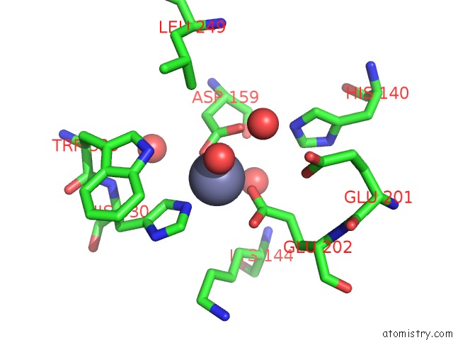
Mono view
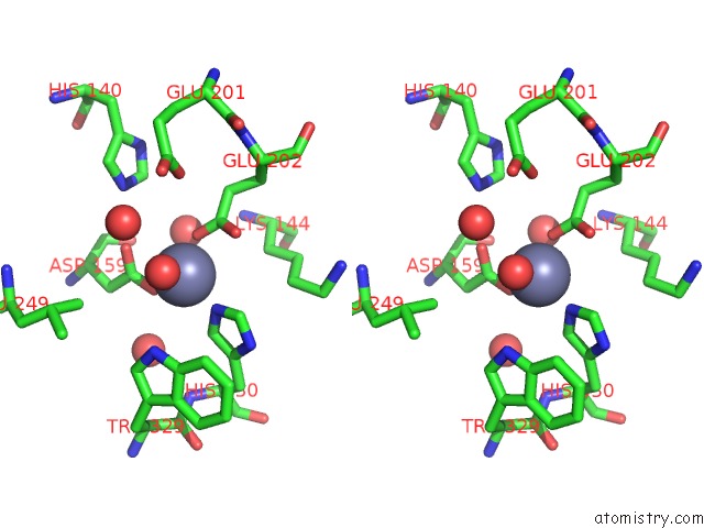
Stereo pair view
Mono view
Stereo pair view
A full contact list of Zinc with other atoms in the Zn binding
site number 1 of Crystal Structure of Double Mutant Y115E Y117E Human Secretory Glutaminyl Cyclase within 5.0Å range:
|
Zinc binding site 2 out of 3 in 7cp0
Go back to
Zinc binding site 2 out
of 3 in the Crystal Structure of Double Mutant Y115E Y117E Human Secretory Glutaminyl Cyclase
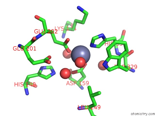
Mono view
Stereo pair view
Mono view
Stereo pair view
A full contact list of Zinc with other atoms in the Zn binding
site number 2 of Crystal Structure of Double Mutant Y115E Y117E Human Secretory Glutaminyl Cyclase within 5.0Å range:
|
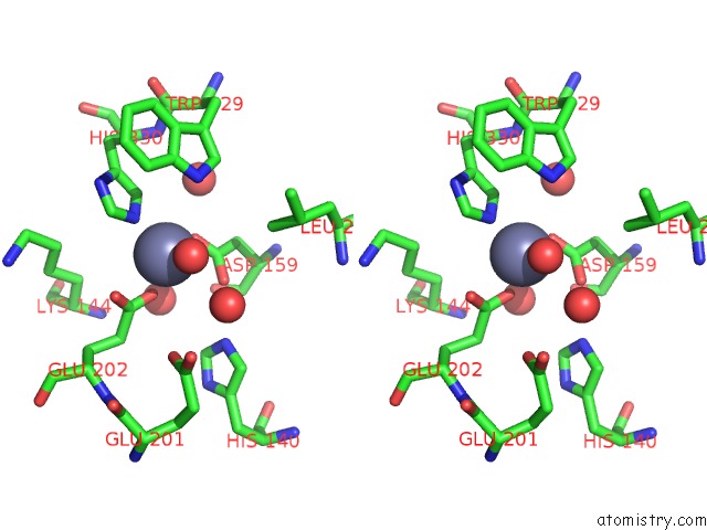
Zinc binding site 3 out of 3 in 7cp0
Go back to
Zinc binding site 3 out
of 3 in the Crystal Structure of Double Mutant Y115E Y117E Human Secretory Glutaminyl Cyclase

Mono view

Stereo pair view
Mono view
Stereo pair view
A full contact list of Zinc with other atoms in the Zn binding
site number 3 of Crystal Structure of Double Mutant Y115E Y117E Human Secretory Glutaminyl Cyclase within 5.0Å range:
|
Reference:
K.V.Dileep,
N.Sakai,
K.Ihara,
M.Kato-Murayama,
A.Nakata,
A.Ito,
D.M.Sivaraman,
J.W.Shin,
M.Yoshida,
M.Shirouzu,
K.Y.J.Zhang.
Piperidine-4-Carboxamide As A New Scaffold For Designing Secretory Glutaminyl Cyclase Inhibitors. Int.J.Biol.Macromol. V. 170 415 2020.
ISSN: ISSN 0141-8130
PubMed: 33373636
DOI: 10.1016/J.IJBIOMAC.2020.12.118
Page generated: Tue Oct 29 18:21:12 2024
ISSN: ISSN 0141-8130
PubMed: 33373636
DOI: 10.1016/J.IJBIOMAC.2020.12.118
Last articles
Mg in 6ZECMg in 6ZE5
Mg in 6ZCJ
Mg in 6ZE4
Mg in 6ZE3
Mg in 6ZE2
Mg in 6ZCE
Mg in 6ZCZ
Mg in 6ZBS
Mg in 6Z9T