Zinc »
PDB 6t6r-6tm5 »
6tld »
Zinc in PDB 6tld: Crystal Structure of Schistosoma Mansoni HDAC8 Complexed with A Triazole Hydroxamate Inhibitor 2
Protein crystallography data
The structure of Crystal Structure of Schistosoma Mansoni HDAC8 Complexed with A Triazole Hydroxamate Inhibitor 2, PDB code: 6tld
was solved by
T.B.Shaik,
C.Romier,
with X-Ray Crystallography technique. A brief refinement statistics is given in the table below:
| Resolution Low / High (Å) | 50.00 / 1.61 |
| Space group | P 1 |
| Cell size a, b, c (Å), α, β, γ (°) | 70.640, 70.720, 98.050, 78.08, 75.69, 85.72 |
| R / Rfree (%) | 16.6 / 19.5 |
Other elements in 6tld:
The structure of Crystal Structure of Schistosoma Mansoni HDAC8 Complexed with A Triazole Hydroxamate Inhibitor 2 also contains other interesting chemical elements:
| Potassium | (K) | 8 atoms |
Zinc Binding Sites:
The binding sites of Zinc atom in the Crystal Structure of Schistosoma Mansoni HDAC8 Complexed with A Triazole Hydroxamate Inhibitor 2
(pdb code 6tld). This binding sites where shown within
5.0 Angstroms radius around Zinc atom.
In total 4 binding sites of Zinc where determined in the Crystal Structure of Schistosoma Mansoni HDAC8 Complexed with A Triazole Hydroxamate Inhibitor 2, PDB code: 6tld:
Jump to Zinc binding site number: 1; 2; 3; 4;
In total 4 binding sites of Zinc where determined in the Crystal Structure of Schistosoma Mansoni HDAC8 Complexed with A Triazole Hydroxamate Inhibitor 2, PDB code: 6tld:
Jump to Zinc binding site number: 1; 2; 3; 4;
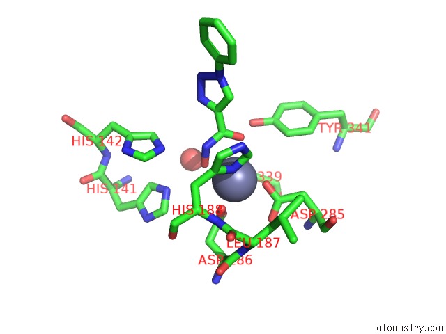
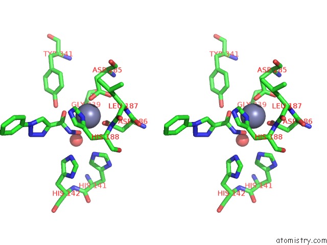
Zinc binding site 1 out of 4 in 6tld
Go back to
Zinc binding site 1 out
of 4 in the Crystal Structure of Schistosoma Mansoni HDAC8 Complexed with A Triazole Hydroxamate Inhibitor 2
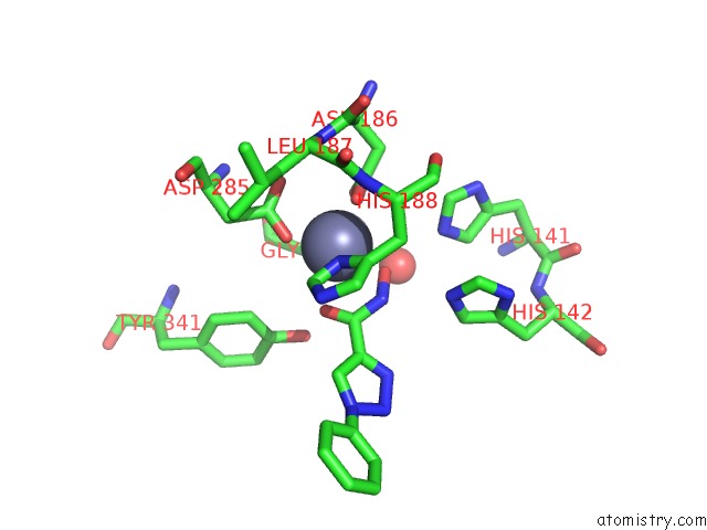
Mono view
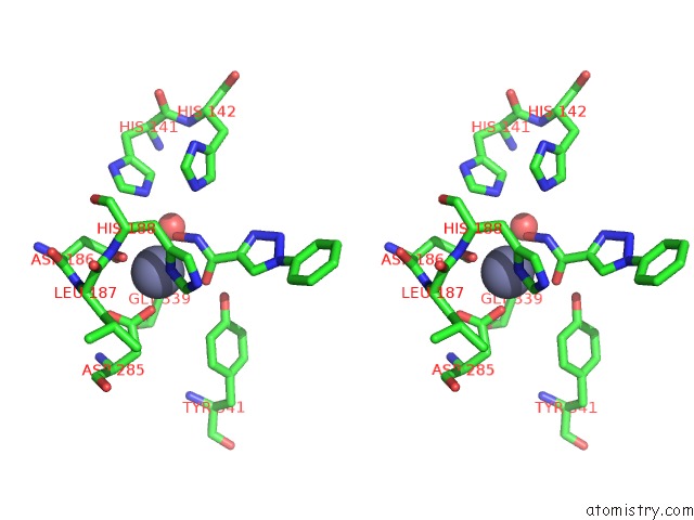
Stereo pair view
Mono view
Stereo pair view
A full contact list of Zinc with other atoms in the Zn binding
site number 1 of Crystal Structure of Schistosoma Mansoni HDAC8 Complexed with A Triazole Hydroxamate Inhibitor 2 within 5.0Å range:
|
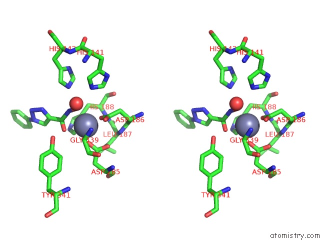
Zinc binding site 2 out of 4 in 6tld
Go back to
Zinc binding site 2 out
of 4 in the Crystal Structure of Schistosoma Mansoni HDAC8 Complexed with A Triazole Hydroxamate Inhibitor 2
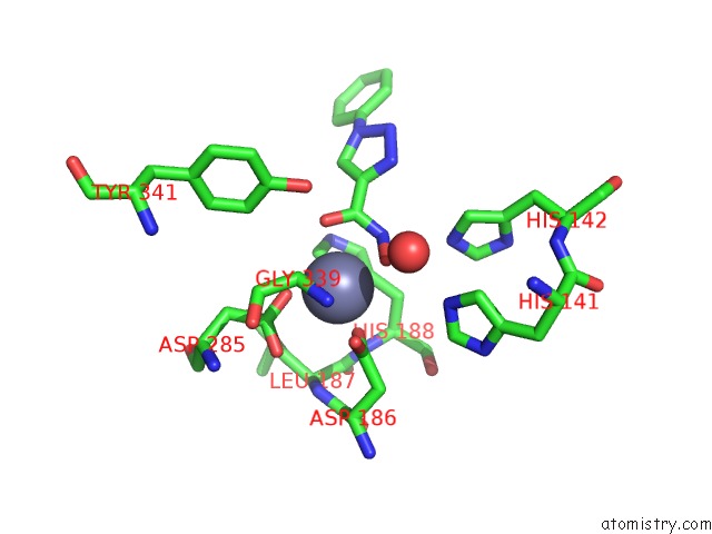
Mono view
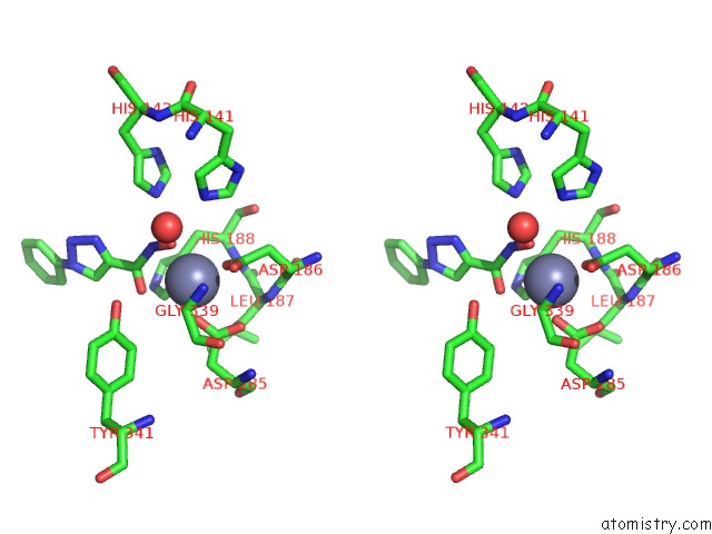
Stereo pair view
Mono view
Stereo pair view
A full contact list of Zinc with other atoms in the Zn binding
site number 2 of Crystal Structure of Schistosoma Mansoni HDAC8 Complexed with A Triazole Hydroxamate Inhibitor 2 within 5.0Å range:
|
Zinc binding site 3 out of 4 in 6tld
Go back to
Zinc binding site 3 out
of 4 in the Crystal Structure of Schistosoma Mansoni HDAC8 Complexed with A Triazole Hydroxamate Inhibitor 2
Mono view
Stereo pair view
Mono view
Stereo pair view
A full contact list of Zinc with other atoms in the Zn binding
site number 3 of Crystal Structure of Schistosoma Mansoni HDAC8 Complexed with A Triazole Hydroxamate Inhibitor 2 within 5.0Å range:
|
Zinc binding site 4 out of 4 in 6tld
Go back to
Zinc binding site 4 out
of 4 in the Crystal Structure of Schistosoma Mansoni HDAC8 Complexed with A Triazole Hydroxamate Inhibitor 2

Mono view
Stereo pair view
Mono view
Stereo pair view
A full contact list of Zinc with other atoms in the Zn binding
site number 4 of Crystal Structure of Schistosoma Mansoni HDAC8 Complexed with A Triazole Hydroxamate Inhibitor 2 within 5.0Å range:
|
Reference:
R.Holl,
D.V.Kalinin,
S.K.Jana,
M.Pfafenrot,
A.Chakrabarti,
J.Melesina,
T.B.Shaik,
J.Lancelot,
R.J.Pierce,
W.Sippl,
C.Romier,
M.Jung.
Structure-Based Design, Synthesis, and Biological Evaluation of Triazole-Based SMHDAC8 Inhibitors. Chemmedchem 2019.
ISSN: ESSN 1860-7187
PubMed: 31816172
DOI: 10.1002/CMDC.201900583
Page generated: Tue Oct 29 08:01:42 2024
ISSN: ESSN 1860-7187
PubMed: 31816172
DOI: 10.1002/CMDC.201900583
Last articles
Mg in 3BM4Mg in 3BL5
Mg in 3BNY
Mg in 3BNX
Mg in 3BN3
Mg in 3BLQ
Mg in 3BJT
Mg in 3BK7
Mg in 3BJF
Mg in 3BK5