Zinc »
PDB 6rpn-6rwn »
6rv5 »
Zinc in PDB 6rv5: X-Ray Structure of the Levansucrase From Erwinia Tasmaniensis in Complex with Levanbiose
Enzymatic activity of X-Ray Structure of the Levansucrase From Erwinia Tasmaniensis in Complex with Levanbiose
All present enzymatic activity of X-Ray Structure of the Levansucrase From Erwinia Tasmaniensis in Complex with Levanbiose:
2.4.1.10;
2.4.1.10;
Protein crystallography data
The structure of X-Ray Structure of the Levansucrase From Erwinia Tasmaniensis in Complex with Levanbiose, PDB code: 6rv5
was solved by
I.Polsinelli,
R.Caliandro,
N.Demitri,
S.Benini,
with X-Ray Crystallography technique. A brief refinement statistics is given in the table below:
| Resolution Low / High (Å) | 45.26 / 1.58 |
| Space group | P 41 21 2 |
| Cell size a, b, c (Å), α, β, γ (°) | 127.886, 127.886, 58.268, 90.00, 90.00, 90.00 |
| R / Rfree (%) | 12.3 / 18.4 |
Zinc Binding Sites:
The binding sites of Zinc atom in the X-Ray Structure of the Levansucrase From Erwinia Tasmaniensis in Complex with Levanbiose
(pdb code 6rv5). This binding sites where shown within
5.0 Angstroms radius around Zinc atom.
In total 2 binding sites of Zinc where determined in the X-Ray Structure of the Levansucrase From Erwinia Tasmaniensis in Complex with Levanbiose, PDB code: 6rv5:
Jump to Zinc binding site number: 1; 2;
In total 2 binding sites of Zinc where determined in the X-Ray Structure of the Levansucrase From Erwinia Tasmaniensis in Complex with Levanbiose, PDB code: 6rv5:
Jump to Zinc binding site number: 1; 2;
Zinc binding site 1 out of 2 in 6rv5
Go back to
Zinc binding site 1 out
of 2 in the X-Ray Structure of the Levansucrase From Erwinia Tasmaniensis in Complex with Levanbiose
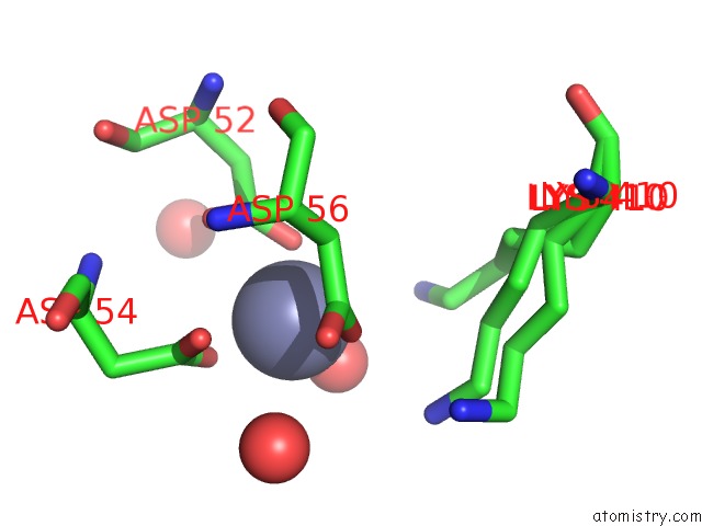
Mono view
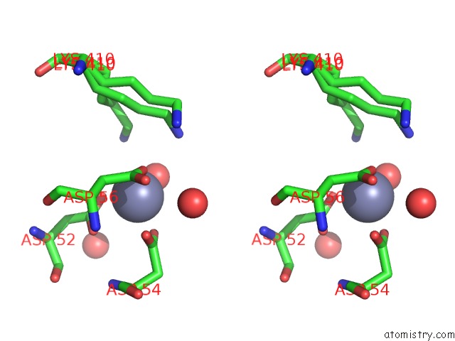
Stereo pair view
Mono view
Stereo pair view
A full contact list of Zinc with other atoms in the Zn binding
site number 1 of X-Ray Structure of the Levansucrase From Erwinia Tasmaniensis in Complex with Levanbiose within 5.0Å range:
|
Zinc binding site 2 out of 2 in 6rv5
Go back to
Zinc binding site 2 out
of 2 in the X-Ray Structure of the Levansucrase From Erwinia Tasmaniensis in Complex with Levanbiose
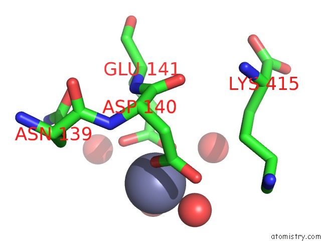
Mono view
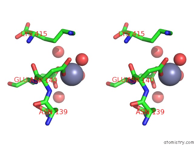
Stereo pair view
Mono view
Stereo pair view
A full contact list of Zinc with other atoms in the Zn binding
site number 2 of X-Ray Structure of the Levansucrase From Erwinia Tasmaniensis in Complex with Levanbiose within 5.0Å range:
|
Reference:
I.Polsinelli,
R.Caliandro,
N.Demitri,
S.Benini.
The Structure of Sucrose-Soaked Levansucrase Crystals From Erwinia Tasmaniensis Reveals A Binding Pocket For Levanbiose Int J Mol Sci 2020.
ISSN: ESSN 1422-0067
Page generated: Tue Oct 29 06:46:48 2024
ISSN: ESSN 1422-0067
Last articles
Mg in 6HNQMg in 6HOS
Mg in 6HNS
Mg in 6HN2
Mg in 6HMZ
Mg in 6HMU
Mg in 6HMT
Mg in 6HLR
Mg in 6HLQ
Mg in 6HKY