Zinc »
PDB 6rg4-6rpc »
6rmr »
Zinc in PDB 6rmr: Crystal Structure of Escherichia Coli Periplasmic Glucose-1- Phosphatase H18D Mutant
Enzymatic activity of Crystal Structure of Escherichia Coli Periplasmic Glucose-1- Phosphatase H18D Mutant
All present enzymatic activity of Crystal Structure of Escherichia Coli Periplasmic Glucose-1- Phosphatase H18D Mutant:
3.1.3.10;
3.1.3.10;
Protein crystallography data
The structure of Crystal Structure of Escherichia Coli Periplasmic Glucose-1- Phosphatase H18D Mutant, PDB code: 6rmr
was solved by
P.Pfeiffer,
G.Oberdorfer,
B.Nidetzky,
with X-Ray Crystallography technique. A brief refinement statistics is given in the table below:
| Resolution Low / High (Å) | 42.77 / 2.50 |
| Space group | P 21 21 21 |
| Cell size a, b, c (Å), α, β, γ (°) | 64.035, 101.651, 114.929, 90.00, 90.00, 90.00 |
| R / Rfree (%) | 22.5 / 26.6 |
Zinc Binding Sites:
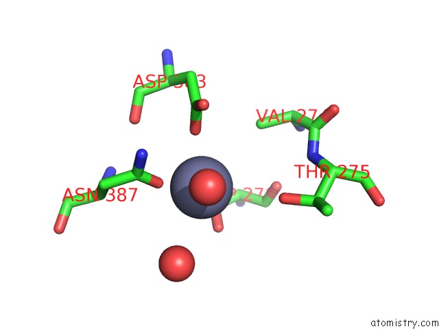
The binding sites of Zinc atom in the Crystal Structure of Escherichia Coli Periplasmic Glucose-1- Phosphatase H18D Mutant
(pdb code 6rmr). This binding sites where shown within
5.0 Angstroms radius around Zinc atom.
In total only one binding site of Zinc was determined in the Crystal Structure of Escherichia Coli Periplasmic Glucose-1- Phosphatase H18D Mutant, PDB code: 6rmr:
In total only one binding site of Zinc was determined in the Crystal Structure of Escherichia Coli Periplasmic Glucose-1- Phosphatase H18D Mutant, PDB code: 6rmr:
Zinc binding site 1 out of 1 in 6rmr
Go back to
Zinc binding site 1 out
of 1 in the Crystal Structure of Escherichia Coli Periplasmic Glucose-1- Phosphatase H18D Mutant
Mono view

Stereo pair view
Mono view
Stereo pair view
A full contact list of Zinc with other atoms in the Zn binding
site number 1 of Crystal Structure of Escherichia Coli Periplasmic Glucose-1- Phosphatase H18D Mutant within 5.0Å range:
|
Reference:
P.Pfeiffer,
G.Oberdorfer,
B.Nidetzky.
Crystal Structure of Escherichia Coli Periplasmic Glucose-1-Phosphatase H18D Mutant To Be Published.
Page generated: Tue Oct 29 06:38:58 2024
Last articles
Fe in 2YXOFe in 2YRS
Fe in 2YXC
Fe in 2YNM
Fe in 2YVJ
Fe in 2YP1
Fe in 2YU2
Fe in 2YU1
Fe in 2YQB
Fe in 2YOO