Zinc »
PDB 6lvu-6mbv »
6m2i »
Zinc in PDB 6m2i: Structure of the 2-Aminoisobutyric Acid Monooxygenase Hydroxylase
Protein crystallography data
The structure of Structure of the 2-Aminoisobutyric Acid Monooxygenase Hydroxylase, PDB code: 6m2i
was solved by
M.Hibi,
B.Mikami,
J.Ogawa,
with X-Ray Crystallography technique. A brief refinement statistics is given in the table below:
| Resolution Low / High (Å) | 44.99 / 2.45 |
| Space group | C 2 2 21 |
| Cell size a, b, c (Å), α, β, γ (°) | 123.751, 206.684, 91.489, 90, 90, 90 |
| R / Rfree (%) | 18.4 / 23.6 |
Other elements in 6m2i:
The structure of Structure of the 2-Aminoisobutyric Acid Monooxygenase Hydroxylase also contains other interesting chemical elements:
| Iron | (Fe) | 2 atoms |
Zinc Binding Sites:
The binding sites of Zinc atom in the Structure of the 2-Aminoisobutyric Acid Monooxygenase Hydroxylase
(pdb code 6m2i). This binding sites where shown within
5.0 Angstroms radius around Zinc atom.
In total only one binding site of Zinc was determined in the Structure of the 2-Aminoisobutyric Acid Monooxygenase Hydroxylase, PDB code: 6m2i:
In total only one binding site of Zinc was determined in the Structure of the 2-Aminoisobutyric Acid Monooxygenase Hydroxylase, PDB code: 6m2i:
Zinc binding site 1 out of 1 in 6m2i
Go back to
Zinc binding site 1 out
of 1 in the Structure of the 2-Aminoisobutyric Acid Monooxygenase Hydroxylase
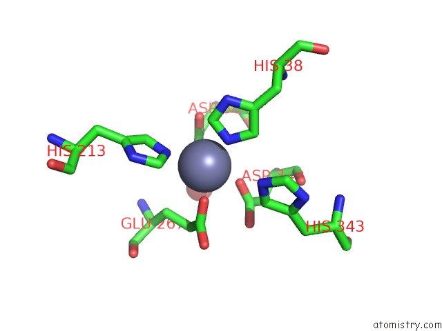
Mono view
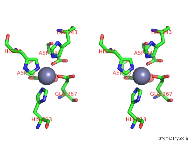
Stereo pair view
Mono view
Stereo pair view
A full contact list of Zinc with other atoms in the Zn binding
site number 1 of Structure of the 2-Aminoisobutyric Acid Monooxygenase Hydroxylase within 5.0Å range:
|
Reference:
M.Hibi,
B.Mikami,
J.Ogawa.
Structure of the 2-Aminoisobutyric Acid Monooxygenase Hydroxylase To Be Published.
Page generated: Tue Oct 29 02:52:37 2024
Last articles
Mg in 7N38Mg in 7N37
Mg in 7N1R
Mg in 7N0Z
Mg in 7N2M
Mg in 7N1O
Mg in 7N0D
Mg in 7N0I
Mg in 7N0C
Mg in 7MZ8