Zinc »
PDB 6j4n-6jeu »
6jeu »
Zinc in PDB 6jeu: K1U Bound Crystal Peptide Deformylase From Acinetobacter Baumanii
Enzymatic activity of K1U Bound Crystal Peptide Deformylase From Acinetobacter Baumanii
All present enzymatic activity of K1U Bound Crystal Peptide Deformylase From Acinetobacter Baumanii:
3.5.1.88;
3.5.1.88;
Protein crystallography data
The structure of K1U Bound Crystal Peptide Deformylase From Acinetobacter Baumanii, PDB code: 6jeu
was solved by
T.H.Ho,
I.H.Lee,
L.W.Kang,
with X-Ray Crystallography technique. A brief refinement statistics is given in the table below:
| Resolution Low / High (Å) | 49.99 / 2.10 |
| Space group | P 32 |
| Cell size a, b, c (Å), α, β, γ (°) | 39.852, 39.852, 187.692, 90.00, 90.00, 120.00 |
| R / Rfree (%) | 21.5 / 28 |
Zinc Binding Sites:
The binding sites of Zinc atom in the K1U Bound Crystal Peptide Deformylase From Acinetobacter Baumanii
(pdb code 6jeu). This binding sites where shown within
5.0 Angstroms radius around Zinc atom.
In total 2 binding sites of Zinc where determined in the K1U Bound Crystal Peptide Deformylase From Acinetobacter Baumanii, PDB code: 6jeu:
Jump to Zinc binding site number: 1; 2;
In total 2 binding sites of Zinc where determined in the K1U Bound Crystal Peptide Deformylase From Acinetobacter Baumanii, PDB code: 6jeu:
Jump to Zinc binding site number: 1; 2;
Zinc binding site 1 out of 2 in 6jeu
Go back to
Zinc binding site 1 out
of 2 in the K1U Bound Crystal Peptide Deformylase From Acinetobacter Baumanii

Mono view
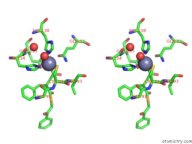
Stereo pair view
Mono view
Stereo pair view
A full contact list of Zinc with other atoms in the Zn binding
site number 1 of K1U Bound Crystal Peptide Deformylase From Acinetobacter Baumanii within 5.0Å range:
|
Zinc binding site 2 out of 2 in 6jeu
Go back to
Zinc binding site 2 out
of 2 in the K1U Bound Crystal Peptide Deformylase From Acinetobacter Baumanii

Mono view

Stereo pair view
Mono view
Stereo pair view
A full contact list of Zinc with other atoms in the Zn binding
site number 2 of K1U Bound Crystal Peptide Deformylase From Acinetobacter Baumanii within 5.0Å range:
|
Reference:
T.H.Ho,
I.H.Lee,
L.W.Kang.
K1U Bound Crystal Peptide Deformylase From Acinetobacter Baumanii To Be Published.
Page generated: Thu Aug 21 16:21:56 2025
Last articles
Zn in 6RXPZn in 6RXS
Zn in 6RXO
Zn in 6RXG
Zn in 6RXL
Zn in 6RXJ
Zn in 6RXK
Zn in 6RWO
Zn in 6RXD
Zn in 6RWN