Zinc »
PDB 6j4f-6jes »
6jde »
Zinc in PDB 6jde: Crystal Structure of A Dna Repair Protein
Enzymatic activity of Crystal Structure of A Dna Repair Protein
All present enzymatic activity of Crystal Structure of A Dna Repair Protein:
3.6.4.12;
3.6.4.12;
Protein crystallography data
The structure of Crystal Structure of A Dna Repair Protein, PDB code: 6jde
was solved by
X.X.Yan,
Q.Tang,
with X-Ray Crystallography technique. A brief refinement statistics is given in the table below:
| Resolution Low / High (Å) | 19.95 / 2.80 |
| Space group | P 1 21 1 |
| Cell size a, b, c (Å), α, β, γ (°) | 80.515, 78.455, 110.587, 90.00, 98.50, 90.00 |
| R / Rfree (%) | 22.8 / 24 |
Zinc Binding Sites:
The binding sites of Zinc atom in the Crystal Structure of A Dna Repair Protein
(pdb code 6jde). This binding sites where shown within
5.0 Angstroms radius around Zinc atom.
In total 2 binding sites of Zinc where determined in the Crystal Structure of A Dna Repair Protein, PDB code: 6jde:
Jump to Zinc binding site number: 1; 2;
In total 2 binding sites of Zinc where determined in the Crystal Structure of A Dna Repair Protein, PDB code: 6jde:
Jump to Zinc binding site number: 1; 2;
Zinc binding site 1 out of 2 in 6jde
Go back to
Zinc binding site 1 out
of 2 in the Crystal Structure of A Dna Repair Protein
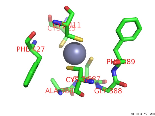
Mono view
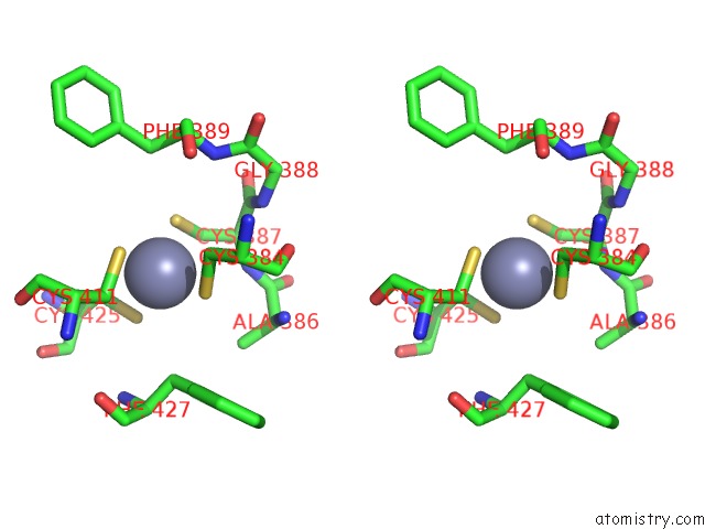
Stereo pair view
Mono view
Stereo pair view
A full contact list of Zinc with other atoms in the Zn binding
site number 1 of Crystal Structure of A Dna Repair Protein within 5.0Å range:
|
Zinc binding site 2 out of 2 in 6jde
Go back to
Zinc binding site 2 out
of 2 in the Crystal Structure of A Dna Repair Protein
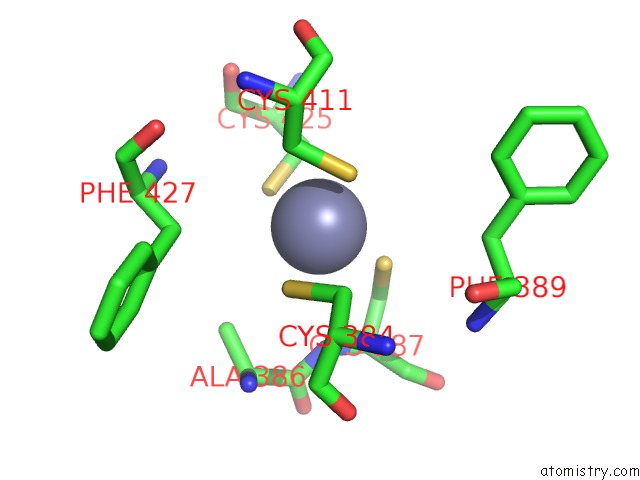
Mono view
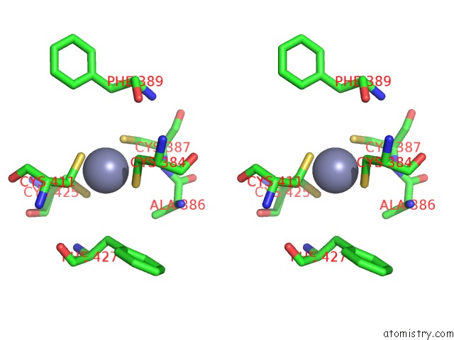
Stereo pair view
Mono view
Stereo pair view
A full contact list of Zinc with other atoms in the Zn binding
site number 2 of Crystal Structure of A Dna Repair Protein within 5.0Å range:
|
Reference:
X.Kuang,
Q.Tang,
Y.P.Liu,
X.X.Yan,
W.Xu.
Crystal Structure of A Novel Atpase Radd From Escherichia Coli. Proteins V. 87 791 2019.
ISSN: ESSN 1097-0134
PubMed: 31035307
DOI: 10.1002/PROT.25704
Page generated: Tue Oct 29 01:02:42 2024
ISSN: ESSN 1097-0134
PubMed: 31035307
DOI: 10.1002/PROT.25704
Last articles
Fe in 2YXOFe in 2YRS
Fe in 2YXC
Fe in 2YNM
Fe in 2YVJ
Fe in 2YP1
Fe in 2YU2
Fe in 2YU1
Fe in 2YQB
Fe in 2YOO