Zinc »
PDB 6i0w-6iiw »
6ie0 »
Zinc in PDB 6ie0: X-Ray Crystal Structure of 2R,3R-Butanediol Dehydrogenase From Bacillus Subtilis
Enzymatic activity of X-Ray Crystal Structure of 2R,3R-Butanediol Dehydrogenase From Bacillus Subtilis
All present enzymatic activity of X-Ray Crystal Structure of 2R,3R-Butanediol Dehydrogenase From Bacillus Subtilis:
1.1.1.4;
1.1.1.4;
Protein crystallography data
The structure of X-Ray Crystal Structure of 2R,3R-Butanediol Dehydrogenase From Bacillus Subtilis, PDB code: 6ie0
was solved by
X.F.Wang,
Y.B.Feng,
F.L.Ji,
with X-Ray Crystallography technique. A brief refinement statistics is given in the table below:
| Resolution Low / High (Å) | 48.71 / 2.98 |
| Space group | P 41 21 2 |
| Cell size a, b, c (Å), α, β, γ (°) | 200.839, 200.839, 83.266, 90.00, 90.00, 90.00 |
| R / Rfree (%) | 17.2 / 22.8 |
Zinc Binding Sites:
The binding sites of Zinc atom in the X-Ray Crystal Structure of 2R,3R-Butanediol Dehydrogenase From Bacillus Subtilis
(pdb code 6ie0). This binding sites where shown within
5.0 Angstroms radius around Zinc atom.
In total 4 binding sites of Zinc where determined in the X-Ray Crystal Structure of 2R,3R-Butanediol Dehydrogenase From Bacillus Subtilis, PDB code: 6ie0:
Jump to Zinc binding site number: 1; 2; 3; 4;
In total 4 binding sites of Zinc where determined in the X-Ray Crystal Structure of 2R,3R-Butanediol Dehydrogenase From Bacillus Subtilis, PDB code: 6ie0:
Jump to Zinc binding site number: 1; 2; 3; 4;
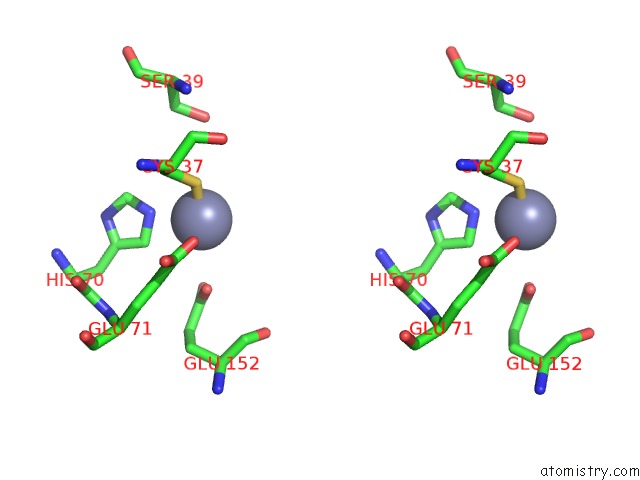
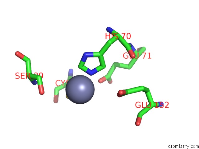
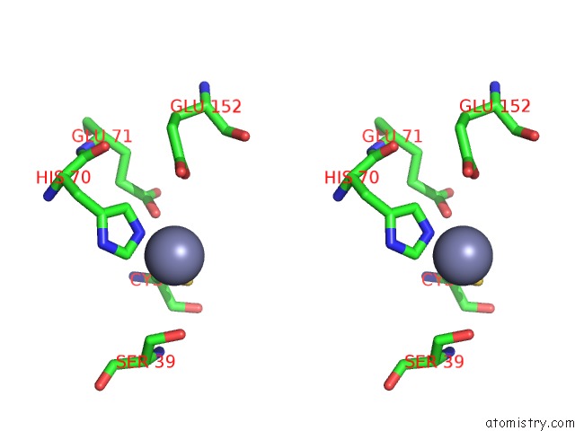
Zinc binding site 1 out of 4 in 6ie0
Go back to
Zinc binding site 1 out
of 4 in the X-Ray Crystal Structure of 2R,3R-Butanediol Dehydrogenase From Bacillus Subtilis

Mono view
Stereo pair view
Mono view
Stereo pair view
A full contact list of Zinc with other atoms in the Zn binding
site number 1 of X-Ray Crystal Structure of 2R,3R-Butanediol Dehydrogenase From Bacillus Subtilis within 5.0Å range:
|
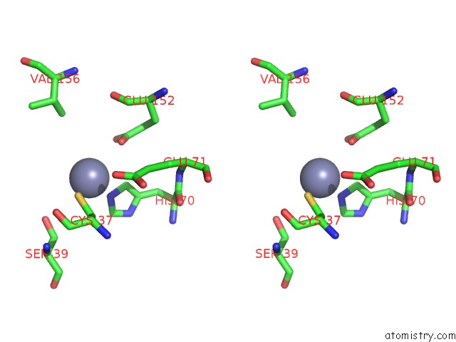
Zinc binding site 2 out of 4 in 6ie0
Go back to
Zinc binding site 2 out
of 4 in the X-Ray Crystal Structure of 2R,3R-Butanediol Dehydrogenase From Bacillus Subtilis

Mono view
Stereo pair view
Mono view
Stereo pair view
A full contact list of Zinc with other atoms in the Zn binding
site number 2 of X-Ray Crystal Structure of 2R,3R-Butanediol Dehydrogenase From Bacillus Subtilis within 5.0Å range:
|
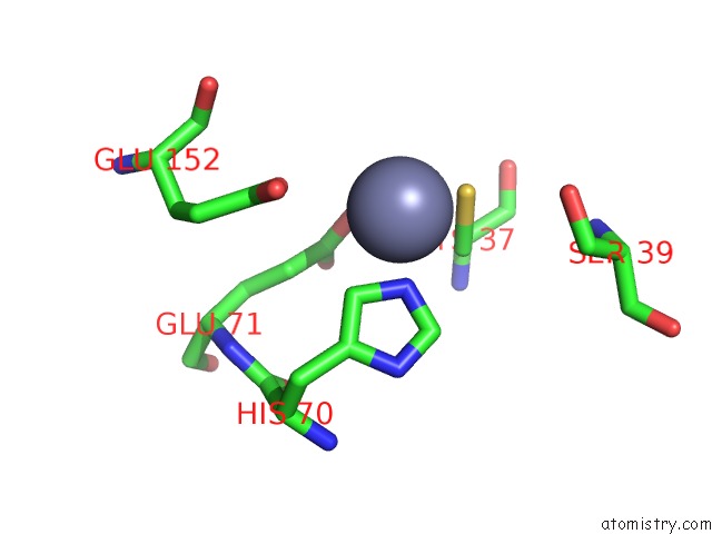
Zinc binding site 3 out of 4 in 6ie0
Go back to
Zinc binding site 3 out
of 4 in the X-Ray Crystal Structure of 2R,3R-Butanediol Dehydrogenase From Bacillus Subtilis
Mono view
Stereo pair view
Mono view
Stereo pair view
A full contact list of Zinc with other atoms in the Zn binding
site number 3 of X-Ray Crystal Structure of 2R,3R-Butanediol Dehydrogenase From Bacillus Subtilis within 5.0Å range:
|
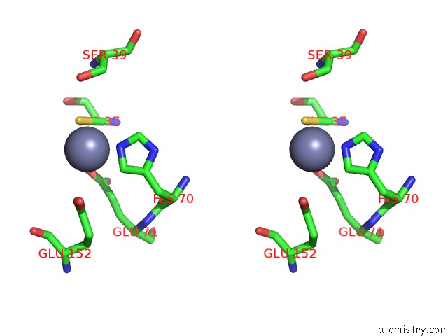
Zinc binding site 4 out of 4 in 6ie0
Go back to
Zinc binding site 4 out
of 4 in the X-Ray Crystal Structure of 2R,3R-Butanediol Dehydrogenase From Bacillus Subtilis
Mono view
Stereo pair view
Mono view
Stereo pair view
A full contact list of Zinc with other atoms in the Zn binding
site number 4 of X-Ray Crystal Structure of 2R,3R-Butanediol Dehydrogenase From Bacillus Subtilis within 5.0Å range:
|
Reference:
X.F.Wang,
Y.B.Feng,
F.L.Ji.
X-Ray Crystal Structure of 2R,3R-Butanediol Dehydrogenase From Bacillus Subtilis To Be Published.
Page generated: Mon Oct 28 23:44:33 2024
Last articles
K in 9GKUK in 9GKX
K in 9ED0
K in 9GEF
K in 9GBY
K in 9G9V
K in 9G9X
K in 9G9W
K in 9FDA
K in 9FCO