Zinc »
PDB 6fso-6g3m »
6fzt »
Zinc in PDB 6fzt: Crystal Structure of SMAD8_9-MH1 Bound to the Ggcgc Site.
Protein crystallography data
The structure of Crystal Structure of SMAD8_9-MH1 Bound to the Ggcgc Site., PDB code: 6fzt
was solved by
Z.Kaczmarska,
J.A.Marquez,
M.J.Macias,
with X-Ray Crystallography technique. A brief refinement statistics is given in the table below:
| Resolution Low / High (Å) | 59.11 / 2.46 |
| Space group | P 21 21 21 |
| Cell size a, b, c (Å), α, β, γ (°) | 75.433, 79.509, 88.372, 90.00, 90.00, 90.00 |
| R / Rfree (%) | 19.3 / 23.7 |
Zinc Binding Sites:
The binding sites of Zinc atom in the Crystal Structure of SMAD8_9-MH1 Bound to the Ggcgc Site.
(pdb code 6fzt). This binding sites where shown within
5.0 Angstroms radius around Zinc atom.
In total 2 binding sites of Zinc where determined in the Crystal Structure of SMAD8_9-MH1 Bound to the Ggcgc Site., PDB code: 6fzt:
Jump to Zinc binding site number: 1; 2;
In total 2 binding sites of Zinc where determined in the Crystal Structure of SMAD8_9-MH1 Bound to the Ggcgc Site., PDB code: 6fzt:
Jump to Zinc binding site number: 1; 2;
Zinc binding site 1 out of 2 in 6fzt
Go back to
Zinc binding site 1 out
of 2 in the Crystal Structure of SMAD8_9-MH1 Bound to the Ggcgc Site.
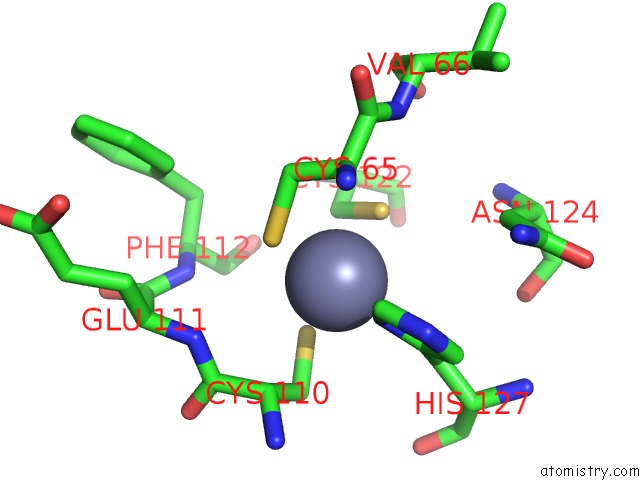
Mono view
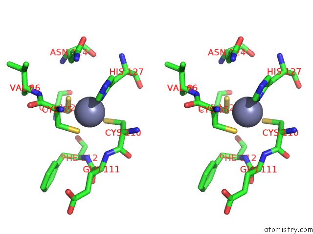
Stereo pair view
Mono view
Stereo pair view
A full contact list of Zinc with other atoms in the Zn binding
site number 1 of Crystal Structure of SMAD8_9-MH1 Bound to the Ggcgc Site. within 5.0Å range:
|
Zinc binding site 2 out of 2 in 6fzt
Go back to
Zinc binding site 2 out
of 2 in the Crystal Structure of SMAD8_9-MH1 Bound to the Ggcgc Site.
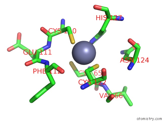
Mono view
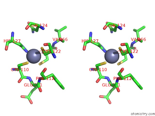
Stereo pair view
Mono view
Stereo pair view
A full contact list of Zinc with other atoms in the Zn binding
site number 2 of Crystal Structure of SMAD8_9-MH1 Bound to the Ggcgc Site. within 5.0Å range:
|
Reference:
L.Ruiz,
Z.Kaczmarska,
E.Aragon,
J.Cordero,
P.Martin Malpartida,
R.Freier,
J.A.Marquez,
M.J.Macias.
Recognition of the 5GC Dna Motif Is Conserved in Bmp and Tgf-Beta Activated Smad Proteins To Be Published.
Page generated: Mon Oct 28 21:33:06 2024
Last articles
Mn in 3GBRMn in 3G1P
Mn in 3G82
Mn in 3GDQ
Mn in 3G3R
Mn in 3G0Z
Mn in 3G10
Mn in 3G2R
Mn in 3G0A
Mn in 3FXO