Zinc »
PDB 6f07-6f98 »
6f6d »
Zinc in PDB 6f6d: The Catalytic Domain of KDM6B in Complex with H3(17-33)K18IA21M Peptide
Protein crystallography data
The structure of The Catalytic Domain of KDM6B in Complex with H3(17-33)K18IA21M Peptide, PDB code: 6f6d
was solved by
S.E.Jones,
L.Olsen,
M.Gajhede,
with X-Ray Crystallography technique. A brief refinement statistics is given in the table below:
| Resolution Low / High (Å) | 57.50 / 1.82 |
| Space group | P 41 21 2 |
| Cell size a, b, c (Å), α, β, γ (°) | 68.480, 68.480, 230.000, 90.00, 90.00, 90.00 |
| R / Rfree (%) | 16.5 / 21.4 |
Other elements in 6f6d:
The structure of The Catalytic Domain of KDM6B in Complex with H3(17-33)K18IA21M Peptide also contains other interesting chemical elements:
| Iron | (Fe) | 1 atom |
Zinc Binding Sites:
The binding sites of Zinc atom in the The Catalytic Domain of KDM6B in Complex with H3(17-33)K18IA21M Peptide
(pdb code 6f6d). This binding sites where shown within
5.0 Angstroms radius around Zinc atom.
In total only one binding site of Zinc was determined in the The Catalytic Domain of KDM6B in Complex with H3(17-33)K18IA21M Peptide, PDB code: 6f6d:
In total only one binding site of Zinc was determined in the The Catalytic Domain of KDM6B in Complex with H3(17-33)K18IA21M Peptide, PDB code: 6f6d:
Zinc binding site 1 out of 1 in 6f6d
Go back to
Zinc binding site 1 out
of 1 in the The Catalytic Domain of KDM6B in Complex with H3(17-33)K18IA21M Peptide
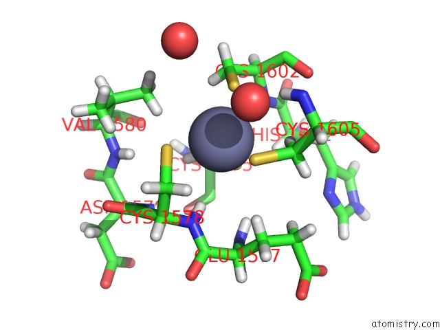
Mono view
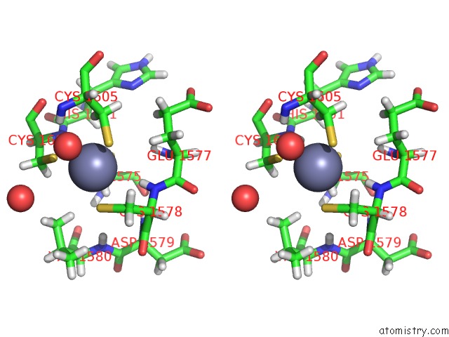
Stereo pair view
Mono view
Stereo pair view
A full contact list of Zinc with other atoms in the Zn binding
site number 1 of The Catalytic Domain of KDM6B in Complex with H3(17-33)K18IA21M Peptide within 5.0Å range:
|
Reference:
S.E.Jones,
L.Olsen,
M.Gajhede.
Structural Basis of Histone Demethylase KDM6B Histone 3 Lysine 27 Specificity. Biochemistry V. 57 585 2018.
ISSN: ISSN 1520-4995
PubMed: 29220567
DOI: 10.1021/ACS.BIOCHEM.7B01152
Page generated: Mon Oct 28 20:44:20 2024
ISSN: ISSN 1520-4995
PubMed: 29220567
DOI: 10.1021/ACS.BIOCHEM.7B01152
Last articles
F in 9VCLF in 9VCK
F in 9JHS
F in 9JF9
F in 9CPK
Cu in 9OH6
Cu in 9OH7
Cu in 9VQM
Cl in 9QM5
Cl in 9RM2