Zinc »
PDB 6div-6dxt »
6dkh »
Zinc in PDB 6dkh: The Crystal Structure of L-Idonate 5-Dehydrogenase From Escherichia Coli Str. K-12 Substr. MG1655
Enzymatic activity of The Crystal Structure of L-Idonate 5-Dehydrogenase From Escherichia Coli Str. K-12 Substr. MG1655
All present enzymatic activity of The Crystal Structure of L-Idonate 5-Dehydrogenase From Escherichia Coli Str. K-12 Substr. MG1655:
1.1.1.264;
1.1.1.264;
Protein crystallography data
The structure of The Crystal Structure of L-Idonate 5-Dehydrogenase From Escherichia Coli Str. K-12 Substr. MG1655, PDB code: 6dkh
was solved by
K.Tan,
E.Evdokimova,
C.Mcchesney,
A.Savchenko,
A.Joachimiak,
Center Forstructural Genomics Of Infectious Diseases (Csgid),
with X-Ray Crystallography technique. A brief refinement statistics is given in the table below:
| Resolution Low / High (Å) | 38.99 / 2.61 |
| Space group | P 1 21 1 |
| Cell size a, b, c (Å), α, β, γ (°) | 78.055, 77.646, 128.350, 90.00, 92.62, 90.00 |
| R / Rfree (%) | 20 / 25.9 |
Zinc Binding Sites:
The binding sites of Zinc atom in the The Crystal Structure of L-Idonate 5-Dehydrogenase From Escherichia Coli Str. K-12 Substr. MG1655
(pdb code 6dkh). This binding sites where shown within
5.0 Angstroms radius around Zinc atom.
In total 4 binding sites of Zinc where determined in the The Crystal Structure of L-Idonate 5-Dehydrogenase From Escherichia Coli Str. K-12 Substr. MG1655, PDB code: 6dkh:
Jump to Zinc binding site number: 1; 2; 3; 4;
In total 4 binding sites of Zinc where determined in the The Crystal Structure of L-Idonate 5-Dehydrogenase From Escherichia Coli Str. K-12 Substr. MG1655, PDB code: 6dkh:
Jump to Zinc binding site number: 1; 2; 3; 4;
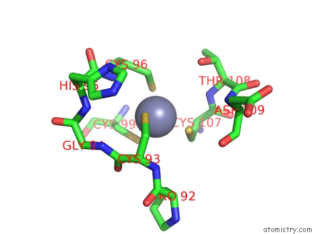
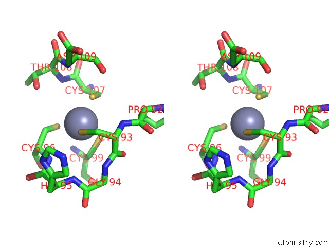
Zinc binding site 1 out of 4 in 6dkh
Go back to
Zinc binding site 1 out
of 4 in the The Crystal Structure of L-Idonate 5-Dehydrogenase From Escherichia Coli Str. K-12 Substr. MG1655
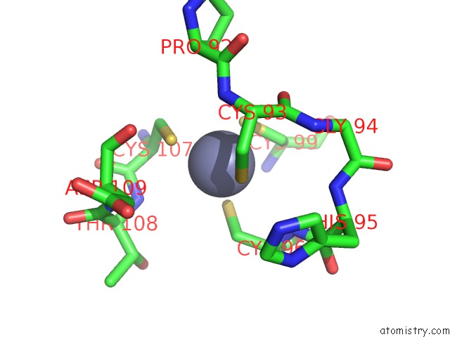
Mono view
Stereo pair view
Mono view
Stereo pair view
A full contact list of Zinc with other atoms in the Zn binding
site number 1 of The Crystal Structure of L-Idonate 5-Dehydrogenase From Escherichia Coli Str. K-12 Substr. MG1655 within 5.0Å range:
|
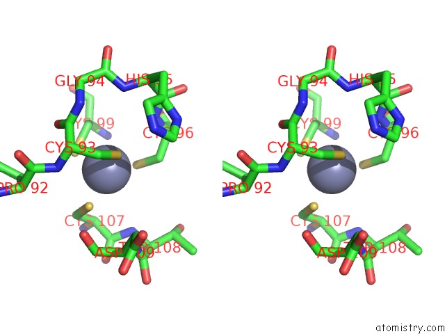
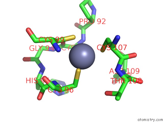
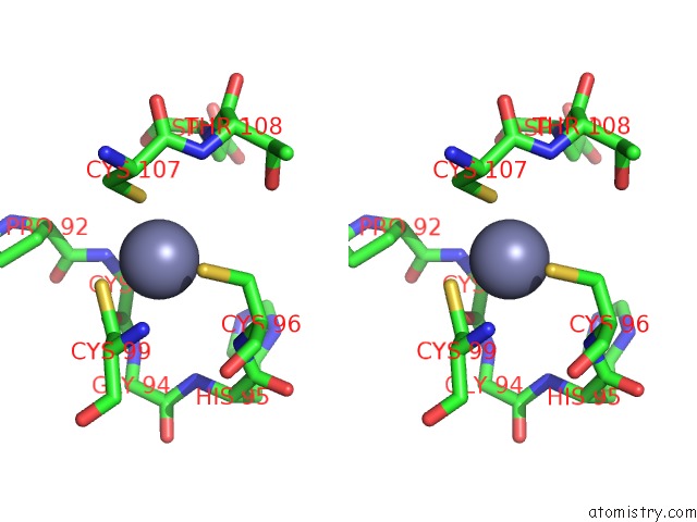
Zinc binding site 2 out of 4 in 6dkh
Go back to
Zinc binding site 2 out
of 4 in the The Crystal Structure of L-Idonate 5-Dehydrogenase From Escherichia Coli Str. K-12 Substr. MG1655
Mono view
Stereo pair view
Mono view
Stereo pair view
A full contact list of Zinc with other atoms in the Zn binding
site number 2 of The Crystal Structure of L-Idonate 5-Dehydrogenase From Escherichia Coli Str. K-12 Substr. MG1655 within 5.0Å range:
|
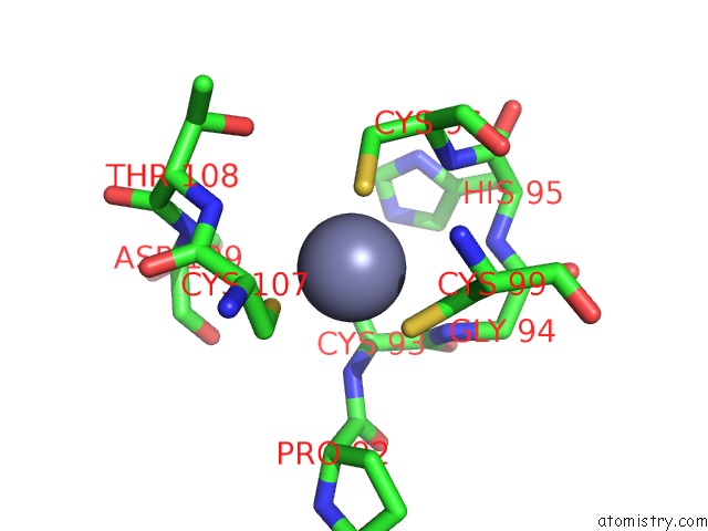
Zinc binding site 3 out of 4 in 6dkh
Go back to
Zinc binding site 3 out
of 4 in the The Crystal Structure of L-Idonate 5-Dehydrogenase From Escherichia Coli Str. K-12 Substr. MG1655
Mono view
Stereo pair view
Mono view
Stereo pair view
A full contact list of Zinc with other atoms in the Zn binding
site number 3 of The Crystal Structure of L-Idonate 5-Dehydrogenase From Escherichia Coli Str. K-12 Substr. MG1655 within 5.0Å range:
|
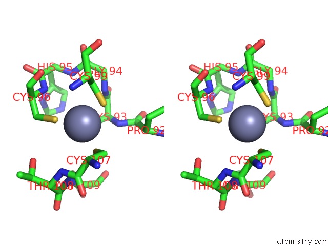
Zinc binding site 4 out of 4 in 6dkh
Go back to
Zinc binding site 4 out
of 4 in the The Crystal Structure of L-Idonate 5-Dehydrogenase From Escherichia Coli Str. K-12 Substr. MG1655
Mono view
Stereo pair view
Mono view
Stereo pair view
A full contact list of Zinc with other atoms in the Zn binding
site number 4 of The Crystal Structure of L-Idonate 5-Dehydrogenase From Escherichia Coli Str. K-12 Substr. MG1655 within 5.0Å range:
|
Reference:
K.Tan,
E.Evdokimova,
C.Mcchesney,
A.Savchenko,
A.Joachimiak.
The Crystal Structure of L-Idonate 5-Dehydrogenase From Escherichia Coli Str. K-12 Substr. MG1655 To Be Published.
Page generated: Mon Oct 28 19:37:56 2024
Last articles
Mg in 6ZXSMg in 7A0Q
Mg in 7A0P
Mg in 7A0C
Mg in 721P
Mg in 6ZXH
Mg in 6ZXG
Mg in 6ZZ6
Mg in 6ZYM
Mg in 6ZY9