Zinc »
PDB 6bto-6c72 »
6bvd »
Zinc in PDB 6bvd: Structure of Botulinum Neurotoxin Serotype Ha Light Chain
Protein crystallography data
The structure of Structure of Botulinum Neurotoxin Serotype Ha Light Chain, PDB code: 6bvd
was solved by
R.Jin,
K.Lam,
with X-Ray Crystallography technique. A brief refinement statistics is given in the table below:
| Resolution Low / High (Å) | 54.05 / 2.09 |
| Space group | C 2 2 21 |
| Cell size a, b, c (Å), α, β, γ (°) | 76.249, 160.745, 219.099, 90.00, 90.00, 90.00 |
| R / Rfree (%) | 17.6 / 19.1 |
Other elements in 6bvd:
The structure of Structure of Botulinum Neurotoxin Serotype Ha Light Chain also contains other interesting chemical elements:
| Calcium | (Ca) | 1 atom |
Zinc Binding Sites:
The binding sites of Zinc atom in the Structure of Botulinum Neurotoxin Serotype Ha Light Chain
(pdb code 6bvd). This binding sites where shown within
5.0 Angstroms radius around Zinc atom.
In total 2 binding sites of Zinc where determined in the Structure of Botulinum Neurotoxin Serotype Ha Light Chain, PDB code: 6bvd:
Jump to Zinc binding site number: 1; 2;
In total 2 binding sites of Zinc where determined in the Structure of Botulinum Neurotoxin Serotype Ha Light Chain, PDB code: 6bvd:
Jump to Zinc binding site number: 1; 2;
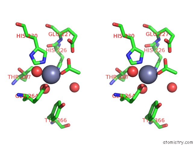
Zinc binding site 1 out of 2 in 6bvd
Go back to
Zinc binding site 1 out
of 2 in the Structure of Botulinum Neurotoxin Serotype Ha Light Chain
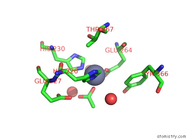
Mono view

Stereo pair view
Mono view
Stereo pair view
A full contact list of Zinc with other atoms in the Zn binding
site number 1 of Structure of Botulinum Neurotoxin Serotype Ha Light Chain within 5.0Å range:
|
Zinc binding site 2 out of 2 in 6bvd
Go back to
Zinc binding site 2 out
of 2 in the Structure of Botulinum Neurotoxin Serotype Ha Light Chain

Mono view
Stereo pair view
Mono view
Stereo pair view
A full contact list of Zinc with other atoms in the Zn binding
site number 2 of Structure of Botulinum Neurotoxin Serotype Ha Light Chain within 5.0Å range:
|
Reference:
K.H.Lam,
S.Sikorra,
J.Weisemann,
H.Maatsch,
K.Perry,
A.Rummel,
T.Binz,
R.Jin.
Structural and Biochemical Characterization of the Protease Domain of the Mosaic Botulinum Neurotoxin Type Ha. Pathog Dis V. 76 2018.
ISSN: ISSN 2049-632X
PubMed: 29688327
DOI: 10.1093/FEMSPD/FTY044
Page generated: Thu Aug 21 12:51:48 2025
ISSN: ISSN 2049-632X
PubMed: 29688327
DOI: 10.1093/FEMSPD/FTY044
Last articles
Mn in 9LJUMn in 9LJW
Mn in 9LJS
Mn in 9LJR
Mn in 9LJT
Mn in 9LJV
Mg in 9UA2
Mg in 9R96
Mg in 9VM1
Mg in 9P01