Zinc »
PDB 5xob-5y1s »
5xrr »
Zinc in PDB 5xrr: Crystal Structure of Fus (54-59) Syssyg
Protein crystallography data
The structure of Crystal Structure of Fus (54-59) Syssyg, PDB code: 5xrr
was solved by
M.Zhao,
X.Gui,
D.Li,
C.Liu,
with X-Ray Crystallography technique. A brief refinement statistics is given in the table below:
| Resolution Low / High (Å) | 20.65 / 1.50 |
| Space group | P 21 21 2 |
| Cell size a, b, c (Å), α, β, γ (°) | 28.240, 30.270, 4.780, 90.00, 90.00, 90.00 |
| R / Rfree (%) | 20.4 / 24.1 |
Zinc Binding Sites:
The binding sites of Zinc atom in the Crystal Structure of Fus (54-59) Syssyg
(pdb code 5xrr). This binding sites where shown within
5.0 Angstroms radius around Zinc atom.
In total only one binding site of Zinc was determined in the Crystal Structure of Fus (54-59) Syssyg, PDB code: 5xrr:
In total only one binding site of Zinc was determined in the Crystal Structure of Fus (54-59) Syssyg, PDB code: 5xrr:
Zinc binding site 1 out of 1 in 5xrr
Go back to
Zinc binding site 1 out
of 1 in the Crystal Structure of Fus (54-59) Syssyg

Mono view
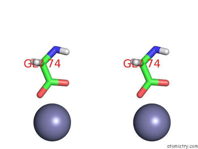
Stereo pair view
Mono view
Stereo pair view
A full contact list of Zinc with other atoms in the Zn binding
site number 1 of Crystal Structure of Fus (54-59) Syssyg within 5.0Å range:
|
Reference:
F.Luo,
X.Gui,
H.Zhou,
J.Gu,
Y.Li,
X.Liu,
M.Zhao,
D.Li,
X.Li,
C.Liu.
Atomic Structures of Fus Lc Domain Segments Reveal Bases For Reversible Amyloid Fibril Formation. Nat. Struct. Mol. Biol. V. 25 341 2018.
ISSN: ESSN 1545-9985
PubMed: 29610493
DOI: 10.1038/S41594-018-0050-8
Page generated: Mon Oct 28 15:13:23 2024
ISSN: ESSN 1545-9985
PubMed: 29610493
DOI: 10.1038/S41594-018-0050-8
Last articles
Fe in 2YXOFe in 2YRS
Fe in 2YXC
Fe in 2YNM
Fe in 2YVJ
Fe in 2YP1
Fe in 2YU2
Fe in 2YU1
Fe in 2YQB
Fe in 2YOO