Zinc »
PDB 5wqa-5x6h »
5wr6 »
Zinc in PDB 5wr6: Thermolysin, Liganded Form with Cryo Condition 2
Enzymatic activity of Thermolysin, Liganded Form with Cryo Condition 2
All present enzymatic activity of Thermolysin, Liganded Form with Cryo Condition 2:
3.4.24.27;
3.4.24.27;
Protein crystallography data
The structure of Thermolysin, Liganded Form with Cryo Condition 2, PDB code: 5wr6
was solved by
N.Kunishima,
H.Naitow,
Y.Matsuura,
with X-Ray Crystallography technique. A brief refinement statistics is given in the table below:
| Resolution Low / High (Å) | 40.20 / 2.30 |
| Space group | P 61 2 2 |
| Cell size a, b, c (Å), α, β, γ (°) | 92.929, 92.929, 129.140, 90.00, 90.00, 120.00 |
| R / Rfree (%) | 15.3 / 18.2 |
Other elements in 5wr6:
The structure of Thermolysin, Liganded Form with Cryo Condition 2 also contains other interesting chemical elements:
| Calcium | (Ca) | 4 atoms |
Zinc Binding Sites:
The binding sites of Zinc atom in the Thermolysin, Liganded Form with Cryo Condition 2
(pdb code 5wr6). This binding sites where shown within
5.0 Angstroms radius around Zinc atom.
In total only one binding site of Zinc was determined in the Thermolysin, Liganded Form with Cryo Condition 2, PDB code: 5wr6:
In total only one binding site of Zinc was determined in the Thermolysin, Liganded Form with Cryo Condition 2, PDB code: 5wr6:
Zinc binding site 1 out of 1 in 5wr6
Go back to
Zinc binding site 1 out
of 1 in the Thermolysin, Liganded Form with Cryo Condition 2
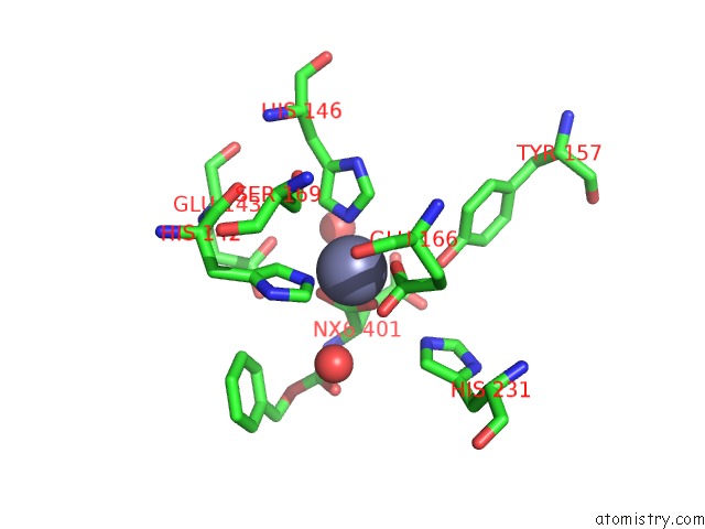
Mono view
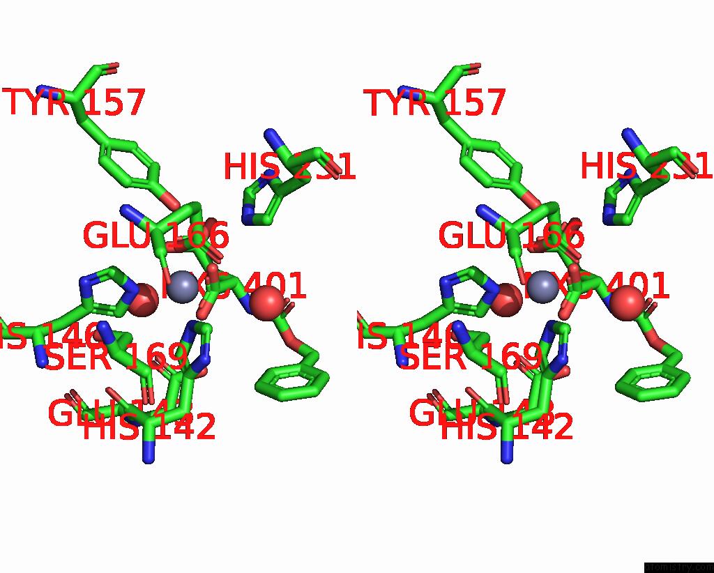
Stereo pair view
Mono view
Stereo pair view
A full contact list of Zinc with other atoms in the Zn binding
site number 1 of Thermolysin, Liganded Form with Cryo Condition 2 within 5.0Å range:
|
Reference:
H.Naitow,
Y.Matsuura,
K.Tono,
Y.Joti,
T.Kameshima,
T.Hatsui,
M.Yabashi,
R.Tanaka,
T.Tanaka,
M.Sugahara,
J.Kobayashi,
E.Nango,
S.Iwata,
N.Kunishima.
Protein-Ligand Complex Structure From Serial Femtosecond Crystallography Using Soaked Thermolysin Microcrystals and Comparison with Structures From Synchrotron Radiation Acta Crystallogr D Struct V. 73 702 2017BIOL.
ISSN: ISSN 2059-7983
PubMed: 28777085
DOI: 10.1107/S2059798317008919
Page generated: Mon Oct 28 14:36:55 2024
ISSN: ISSN 2059-7983
PubMed: 28777085
DOI: 10.1107/S2059798317008919
Last articles
Fe in 4NBHFe in 4NBG
Fe in 4NJB
Fe in 4NI1
Fe in 4NI0
Fe in 4NFG
Fe in 4NED
Fe in 4NE0
Fe in 4NBF
Fe in 4NBC