Zinc »
PDB 5urb-5v3c »
5uxs »
Zinc in PDB 5uxs: X Ray Structure of the Periplasmic Ligand Binding Protein Yfea From Yersinia Pestis
Protein crystallography data
The structure of X Ray Structure of the Periplasmic Ligand Binding Protein Yfea From Yersinia Pestis, PDB code: 5uxs
was solved by
C.D.Radka,
L.J.Delucas,
S.G.Aller,
with X-Ray Crystallography technique. A brief refinement statistics is given in the table below:
| Resolution Low / High (Å) | 33.58 / 1.42 |
| Space group | P 21 21 21 |
| Cell size a, b, c (Å), α, β, γ (°) | 41.736, 52.120, 113.066, 90.00, 90.00, 90.00 |
| R / Rfree (%) | 17.9 / 19 |
Zinc Binding Sites:
The binding sites of Zinc atom in the X Ray Structure of the Periplasmic Ligand Binding Protein Yfea From Yersinia Pestis
(pdb code 5uxs). This binding sites where shown within
5.0 Angstroms radius around Zinc atom.
In total only one binding site of Zinc was determined in the X Ray Structure of the Periplasmic Ligand Binding Protein Yfea From Yersinia Pestis, PDB code: 5uxs:
In total only one binding site of Zinc was determined in the X Ray Structure of the Periplasmic Ligand Binding Protein Yfea From Yersinia Pestis, PDB code: 5uxs:
Zinc binding site 1 out of 1 in 5uxs
Go back to
Zinc binding site 1 out
of 1 in the X Ray Structure of the Periplasmic Ligand Binding Protein Yfea From Yersinia Pestis
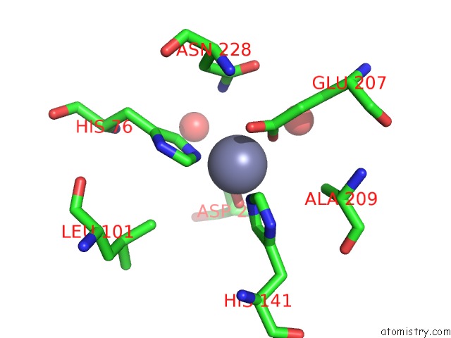
Mono view
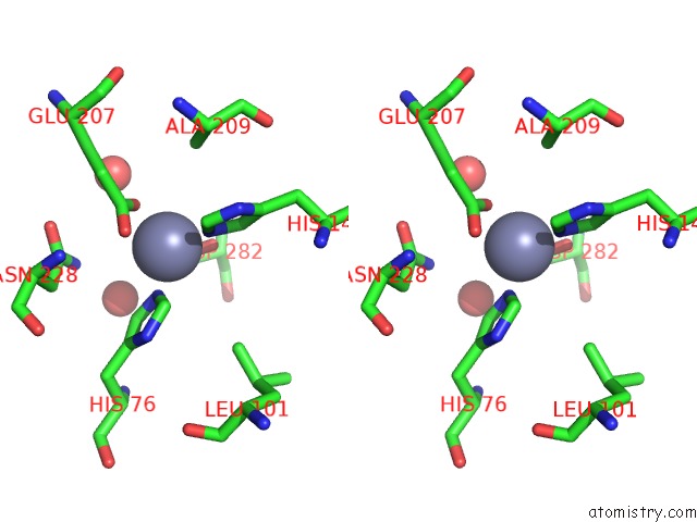
Stereo pair view
Mono view
Stereo pair view
A full contact list of Zinc with other atoms in the Zn binding
site number 1 of X Ray Structure of the Periplasmic Ligand Binding Protein Yfea From Yersinia Pestis within 5.0Å range:
|
Reference:
C.D.Radka,
L.J.Delucas,
L.S.Wilson,
M.B.Lawrenz,
R.D.Perry,
S.G.Aller.
Crystal Structure of Yersinia Pestis Virulence Factor Yfea Reveals Two Polyspecific Metal-Binding Sites. Acta Crystallogr D Struct V. 73 557 2017BIOL.
ISSN: ISSN 2059-7983
PubMed: 28695856
DOI: 10.1107/S2059798317006349
Page generated: Mon Oct 28 12:28:05 2024
ISSN: ISSN 2059-7983
PubMed: 28695856
DOI: 10.1107/S2059798317006349
Last articles
Fe in 2YXOFe in 2YRS
Fe in 2YXC
Fe in 2YNM
Fe in 2YVJ
Fe in 2YP1
Fe in 2YU2
Fe in 2YU1
Fe in 2YQB
Fe in 2YOO