Zinc »
PDB 5urb-5v3c »
5uw2 »
Zinc in PDB 5uw2: Structure of E. Coli Mce Protein Mlad, Periplasmic Domain
Protein crystallography data
The structure of Structure of E. Coli Mce Protein Mlad, Periplasmic Domain, PDB code: 5uw2
was solved by
G.Bhabha,
D.C.Ekiert,
with X-Ray Crystallography technique. A brief refinement statistics is given in the table below:
| Resolution Low / High (Å) | 33.60 / 2.85 |
| Space group | C 1 2 1 |
| Cell size a, b, c (Å), α, β, γ (°) | 122.360, 63.820, 91.150, 90.00, 130.01, 90.00 |
| R / Rfree (%) | 23.9 / 26.5 |
Zinc Binding Sites:
The binding sites of Zinc atom in the Structure of E. Coli Mce Protein Mlad, Periplasmic Domain
(pdb code 5uw2). This binding sites where shown within
5.0 Angstroms radius around Zinc atom.
In total 6 binding sites of Zinc where determined in the Structure of E. Coli Mce Protein Mlad, Periplasmic Domain, PDB code: 5uw2:
Jump to Zinc binding site number: 1; 2; 3; 4; 5; 6;
In total 6 binding sites of Zinc where determined in the Structure of E. Coli Mce Protein Mlad, Periplasmic Domain, PDB code: 5uw2:
Jump to Zinc binding site number: 1; 2; 3; 4; 5; 6;
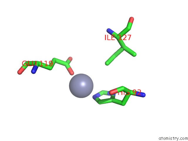
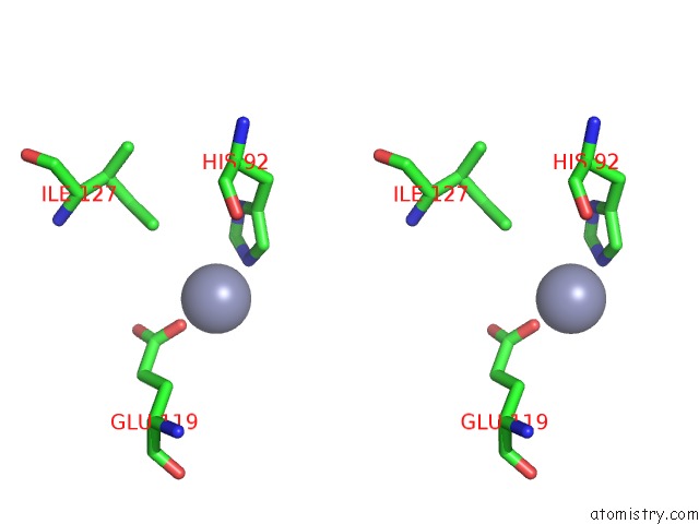
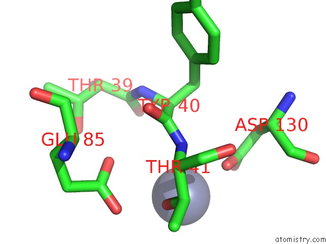
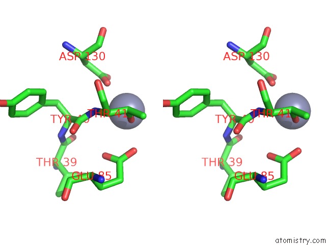
Zinc binding site 1 out of 6 in 5uw2
Go back to
Zinc binding site 1 out
of 6 in the Structure of E. Coli Mce Protein Mlad, Periplasmic Domain
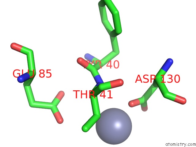
Mono view
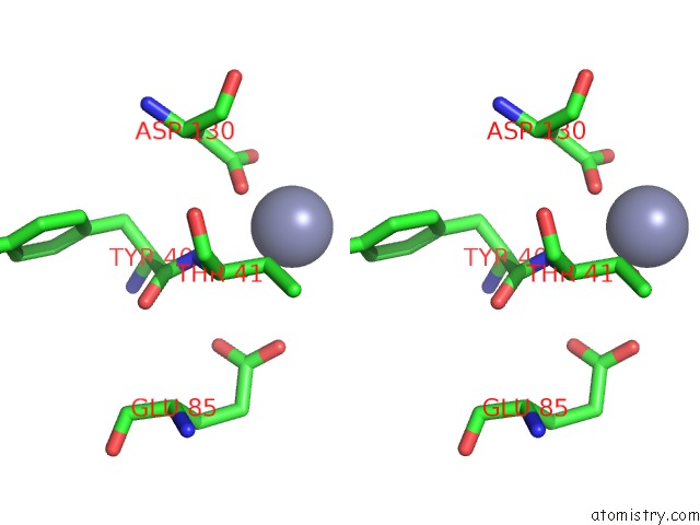
Stereo pair view
Mono view
Stereo pair view
A full contact list of Zinc with other atoms in the Zn binding
site number 1 of Structure of E. Coli Mce Protein Mlad, Periplasmic Domain within 5.0Å range:
|
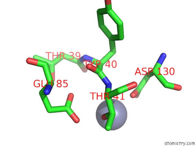
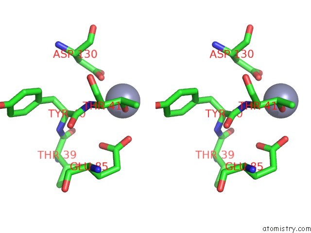
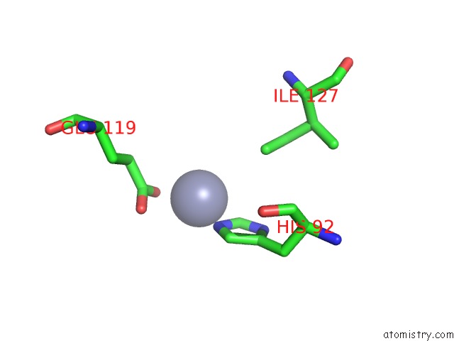
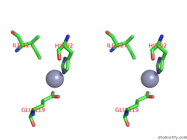
Zinc binding site 2 out of 6 in 5uw2
Go back to
Zinc binding site 2 out
of 6 in the Structure of E. Coli Mce Protein Mlad, Periplasmic Domain
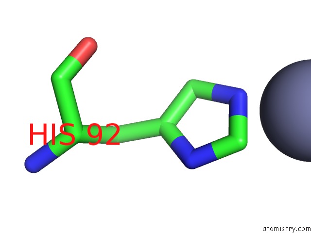
Mono view
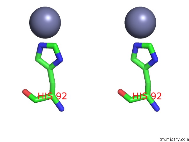
Stereo pair view
Mono view
Stereo pair view
A full contact list of Zinc with other atoms in the Zn binding
site number 2 of Structure of E. Coli Mce Protein Mlad, Periplasmic Domain within 5.0Å range:
|
Zinc binding site 3 out of 6 in 5uw2
Go back to
Zinc binding site 3 out
of 6 in the Structure of E. Coli Mce Protein Mlad, Periplasmic Domain
Mono view
Stereo pair view
Mono view
Stereo pair view
A full contact list of Zinc with other atoms in the Zn binding
site number 3 of Structure of E. Coli Mce Protein Mlad, Periplasmic Domain within 5.0Å range:
|
Zinc binding site 4 out of 6 in 5uw2
Go back to
Zinc binding site 4 out
of 6 in the Structure of E. Coli Mce Protein Mlad, Periplasmic Domain
Mono view
Stereo pair view
Mono view
Stereo pair view
A full contact list of Zinc with other atoms in the Zn binding
site number 4 of Structure of E. Coli Mce Protein Mlad, Periplasmic Domain within 5.0Å range:
|
Zinc binding site 5 out of 6 in 5uw2
Go back to
Zinc binding site 5 out
of 6 in the Structure of E. Coli Mce Protein Mlad, Periplasmic Domain
Mono view
Stereo pair view
Mono view
Stereo pair view
A full contact list of Zinc with other atoms in the Zn binding
site number 5 of Structure of E. Coli Mce Protein Mlad, Periplasmic Domain within 5.0Å range:
|
Zinc binding site 6 out of 6 in 5uw2
Go back to
Zinc binding site 6 out
of 6 in the Structure of E. Coli Mce Protein Mlad, Periplasmic Domain
Mono view
Stereo pair view
Mono view
Stereo pair view
A full contact list of Zinc with other atoms in the Zn binding
site number 6 of Structure of E. Coli Mce Protein Mlad, Periplasmic Domain within 5.0Å range:
|
Reference:
D.C.Ekiert,
G.Bhabha,
G.L.Isom,
G.Greenan,
S.Ovchinnikov,
I.R.Henderson,
J.S.Cox,
R.D.Vale.
Architectures of Lipid Transport Systems For the Bacterial Outer Membrane. Cell V. 169 273 2017.
ISSN: ISSN 1097-4172
PubMed: 28388411
DOI: 10.1016/J.CELL.2017.03.019
Page generated: Mon Oct 28 12:25:30 2024
ISSN: ISSN 1097-4172
PubMed: 28388411
DOI: 10.1016/J.CELL.2017.03.019
Last articles
Fe in 2YXOFe in 2YRS
Fe in 2YXC
Fe in 2YNM
Fe in 2YVJ
Fe in 2YP1
Fe in 2YU2
Fe in 2YU1
Fe in 2YQB
Fe in 2YOO