Zinc »
PDB 5uru-5v3g »
5utj »
Zinc in PDB 5utj: Crystal Structure of Tgt in Complex with 2,6-Dioxy-8-Azapurine, 2,6- Dioxy-8-Azapurine, 2,6-Dioxy-8-Azapurine
Enzymatic activity of Crystal Structure of Tgt in Complex with 2,6-Dioxy-8-Azapurine, 2,6- Dioxy-8-Azapurine, 2,6-Dioxy-8-Azapurine
All present enzymatic activity of Crystal Structure of Tgt in Complex with 2,6-Dioxy-8-Azapurine, 2,6- Dioxy-8-Azapurine, 2,6-Dioxy-8-Azapurine:
2.4.2.29;
2.4.2.29;
Protein crystallography data
The structure of Crystal Structure of Tgt in Complex with 2,6-Dioxy-8-Azapurine, 2,6- Dioxy-8-Azapurine, 2,6-Dioxy-8-Azapurine, PDB code: 5utj
was solved by
E.Hassaan,
A.Heine,
G.Klebe,
with X-Ray Crystallography technique. A brief refinement statistics is given in the table below:
| Resolution Low / High (Å) | 44.50 / 1.55 |
| Space group | C 1 2 1 |
| Cell size a, b, c (Å), α, β, γ (°) | 89.142, 64.759, 70.451, 90.00, 93.15, 90.00 |
| R / Rfree (%) | 14.7 / 17.7 |
Zinc Binding Sites:
The binding sites of Zinc atom in the Crystal Structure of Tgt in Complex with 2,6-Dioxy-8-Azapurine, 2,6- Dioxy-8-Azapurine, 2,6-Dioxy-8-Azapurine
(pdb code 5utj). This binding sites where shown within
5.0 Angstroms radius around Zinc atom.
In total only one binding site of Zinc was determined in the Crystal Structure of Tgt in Complex with 2,6-Dioxy-8-Azapurine, 2,6- Dioxy-8-Azapurine, 2,6-Dioxy-8-Azapurine, PDB code: 5utj:
In total only one binding site of Zinc was determined in the Crystal Structure of Tgt in Complex with 2,6-Dioxy-8-Azapurine, 2,6- Dioxy-8-Azapurine, 2,6-Dioxy-8-Azapurine, PDB code: 5utj:
Zinc binding site 1 out of 1 in 5utj
Go back to
Zinc binding site 1 out
of 1 in the Crystal Structure of Tgt in Complex with 2,6-Dioxy-8-Azapurine, 2,6- Dioxy-8-Azapurine, 2,6-Dioxy-8-Azapurine
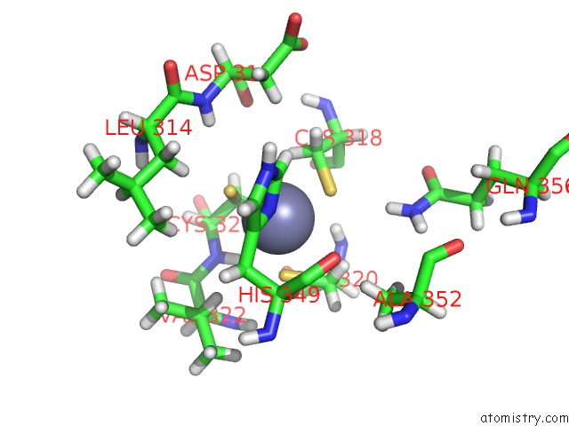
Mono view
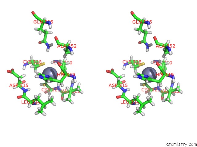
Stereo pair view
Mono view
Stereo pair view
A full contact list of Zinc with other atoms in the Zn binding
site number 1 of Crystal Structure of Tgt in Complex with 2,6-Dioxy-8-Azapurine, 2,6- Dioxy-8-Azapurine, 2,6-Dioxy-8-Azapurine within 5.0Å range:
|
Reference:
E.Hassaan,
A.Heine,
G.Klebe.
Crystal Structure of Tgt in Complex with 2,6-Dioxy-8-Azapurine, 2,6-Dioxy-8-Azapurine, 2,6-Dioxy-8-Azapurine To Be Published.
Page generated: Thu Aug 21 09:37:53 2025
Last articles
Zn in 6F40Zn in 6F3N
Zn in 6F3M
Zn in 6F3O
Zn in 6F3K
Zn in 6F33
Zn in 6F30
Zn in 6F2Y
Zn in 6F3B
Zn in 6F2V