Zinc »
PDB 5t74-5th9 »
5tdc »
Zinc in PDB 5tdc: Crystal Structure of the Human Ubr-Box Domain From UBR1 in Complex with Monomethylated Arginine Peptide.
Protein crystallography data
The structure of Crystal Structure of the Human Ubr-Box Domain From UBR1 in Complex with Monomethylated Arginine Peptide., PDB code: 5tdc
was solved by
G.Kozlov,
J.Munoz-Escobar,
E.Matta-Camacho,
K.Gehring,
with X-Ray Crystallography technique. A brief refinement statistics is given in the table below:
| Resolution Low / High (Å) | 28.74 / 1.61 |
| Space group | P 21 21 21 |
| Cell size a, b, c (Å), α, β, γ (°) | 47.274, 49.036, 53.634, 90.00, 90.00, 90.00 |
| R / Rfree (%) | 14.9 / 18 |
Zinc Binding Sites:
The binding sites of Zinc atom in the Crystal Structure of the Human Ubr-Box Domain From UBR1 in Complex with Monomethylated Arginine Peptide.
(pdb code 5tdc). This binding sites where shown within
5.0 Angstroms radius around Zinc atom.
In total 6 binding sites of Zinc where determined in the Crystal Structure of the Human Ubr-Box Domain From UBR1 in Complex with Monomethylated Arginine Peptide., PDB code: 5tdc:
Jump to Zinc binding site number: 1; 2; 3; 4; 5; 6;
In total 6 binding sites of Zinc where determined in the Crystal Structure of the Human Ubr-Box Domain From UBR1 in Complex with Monomethylated Arginine Peptide., PDB code: 5tdc:
Jump to Zinc binding site number: 1; 2; 3; 4; 5; 6;
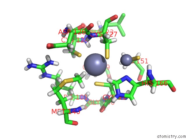
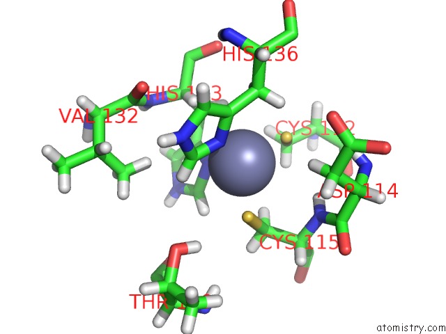
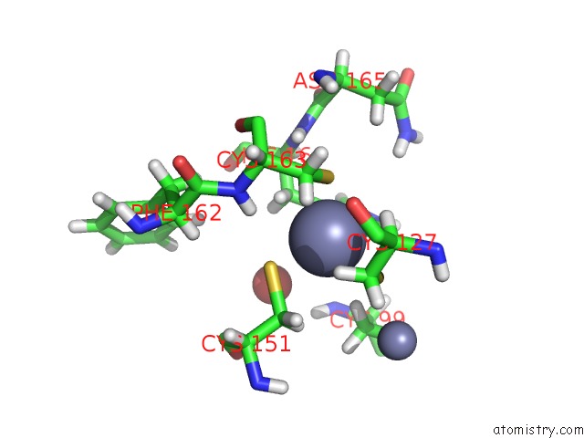
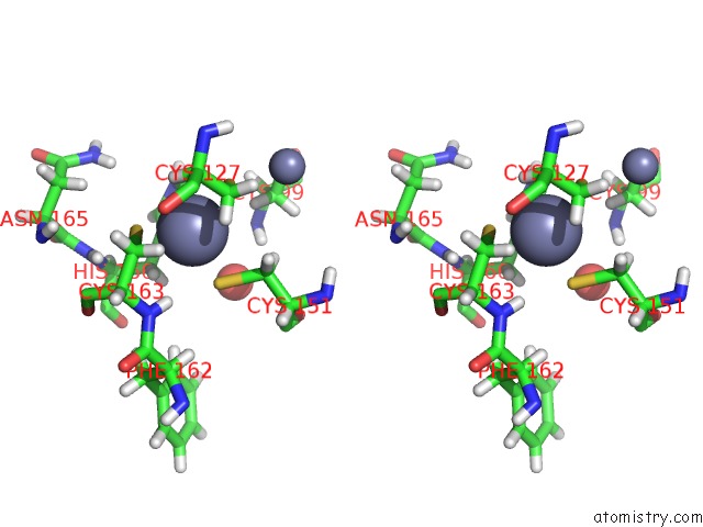
Zinc binding site 1 out of 6 in 5tdc
Go back to
Zinc binding site 1 out
of 6 in the Crystal Structure of the Human Ubr-Box Domain From UBR1 in Complex with Monomethylated Arginine Peptide.
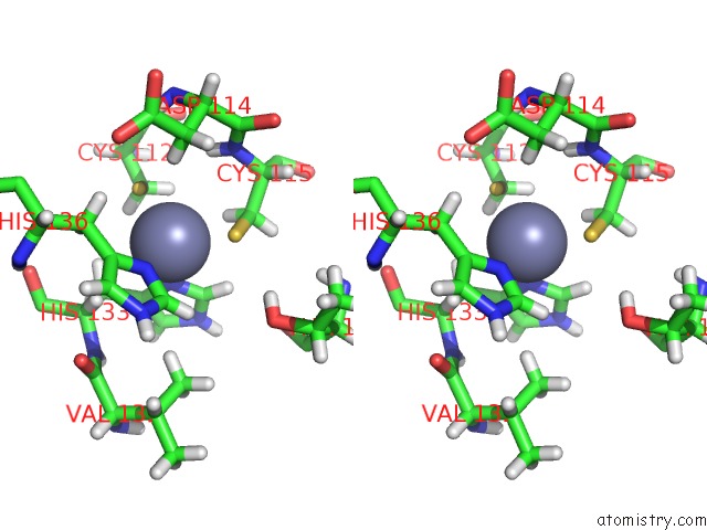
Mono view
Stereo pair view
Mono view
Stereo pair view
A full contact list of Zinc with other atoms in the Zn binding
site number 1 of Crystal Structure of the Human Ubr-Box Domain From UBR1 in Complex with Monomethylated Arginine Peptide. within 5.0Å range:
|
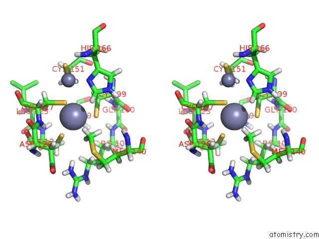
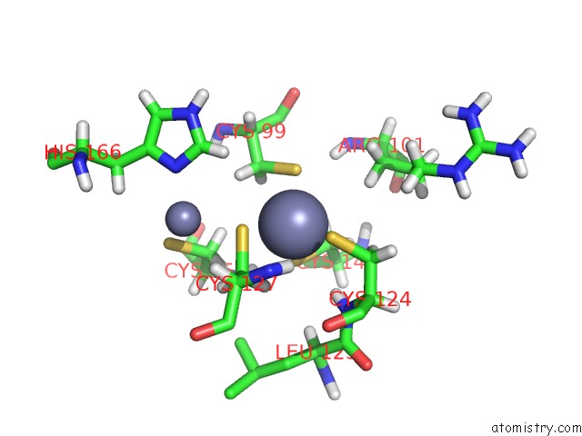
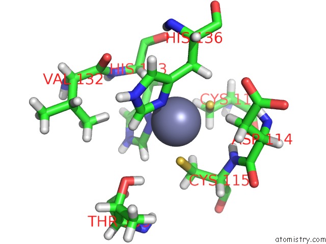
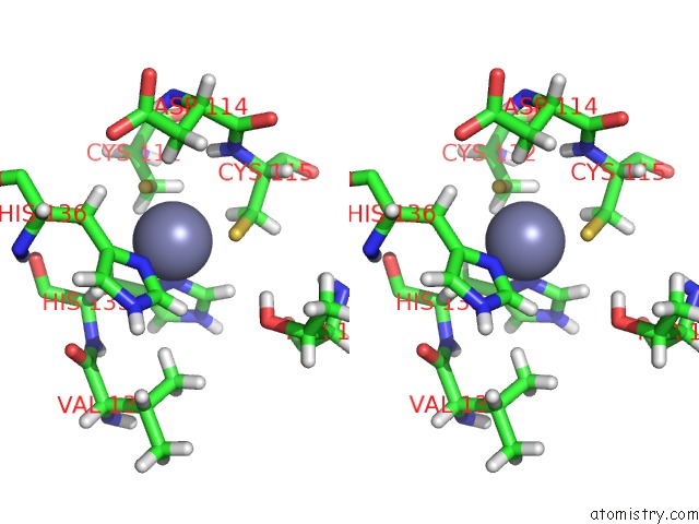
Zinc binding site 2 out of 6 in 5tdc
Go back to
Zinc binding site 2 out
of 6 in the Crystal Structure of the Human Ubr-Box Domain From UBR1 in Complex with Monomethylated Arginine Peptide.
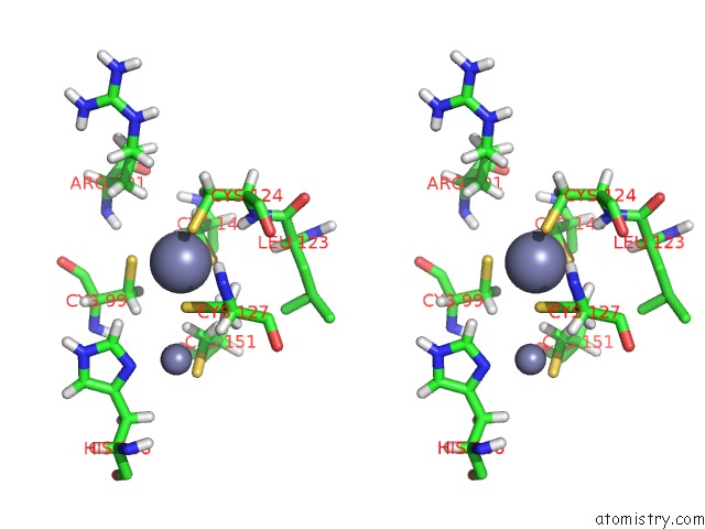
Mono view
Stereo pair view
Mono view
Stereo pair view
A full contact list of Zinc with other atoms in the Zn binding
site number 2 of Crystal Structure of the Human Ubr-Box Domain From UBR1 in Complex with Monomethylated Arginine Peptide. within 5.0Å range:
|
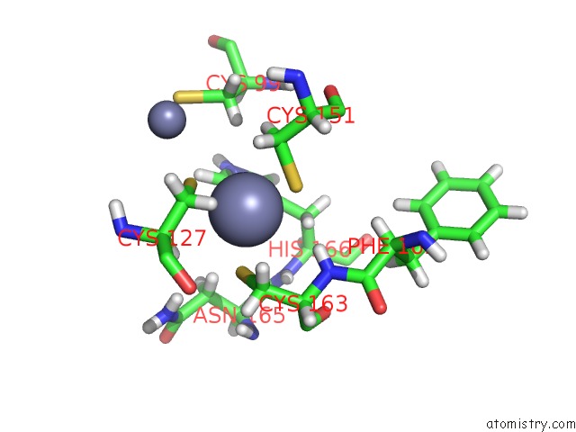
Zinc binding site 3 out of 6 in 5tdc
Go back to
Zinc binding site 3 out
of 6 in the Crystal Structure of the Human Ubr-Box Domain From UBR1 in Complex with Monomethylated Arginine Peptide.
Mono view
Stereo pair view
Mono view
Stereo pair view
A full contact list of Zinc with other atoms in the Zn binding
site number 3 of Crystal Structure of the Human Ubr-Box Domain From UBR1 in Complex with Monomethylated Arginine Peptide. within 5.0Å range:
|
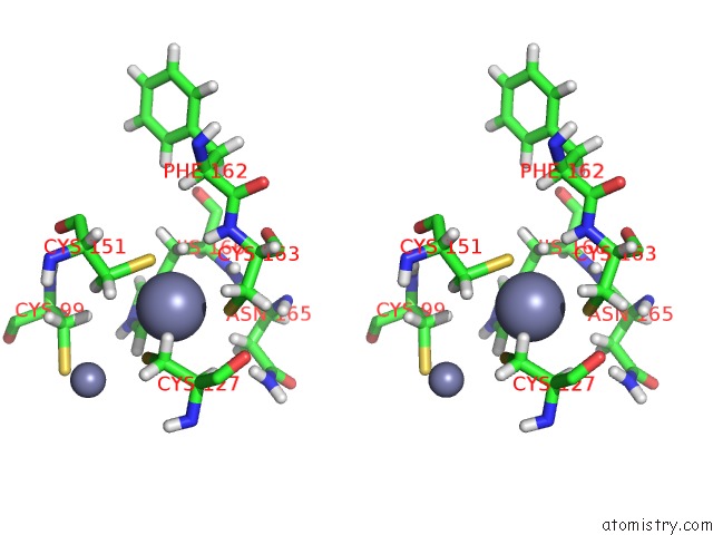
Zinc binding site 4 out of 6 in 5tdc
Go back to
Zinc binding site 4 out
of 6 in the Crystal Structure of the Human Ubr-Box Domain From UBR1 in Complex with Monomethylated Arginine Peptide.
Mono view
Stereo pair view
Mono view
Stereo pair view
A full contact list of Zinc with other atoms in the Zn binding
site number 4 of Crystal Structure of the Human Ubr-Box Domain From UBR1 in Complex with Monomethylated Arginine Peptide. within 5.0Å range:
|
Zinc binding site 5 out of 6 in 5tdc
Go back to
Zinc binding site 5 out
of 6 in the Crystal Structure of the Human Ubr-Box Domain From UBR1 in Complex with Monomethylated Arginine Peptide.
Mono view
Stereo pair view
Mono view
Stereo pair view
A full contact list of Zinc with other atoms in the Zn binding
site number 5 of Crystal Structure of the Human Ubr-Box Domain From UBR1 in Complex with Monomethylated Arginine Peptide. within 5.0Å range:
|
Zinc binding site 6 out of 6 in 5tdc
Go back to
Zinc binding site 6 out
of 6 in the Crystal Structure of the Human Ubr-Box Domain From UBR1 in Complex with Monomethylated Arginine Peptide.
Mono view
Stereo pair view
Mono view
Stereo pair view
A full contact list of Zinc with other atoms in the Zn binding
site number 6 of Crystal Structure of the Human Ubr-Box Domain From UBR1 in Complex with Monomethylated Arginine Peptide. within 5.0Å range:
|
Reference:
J.Munoz-Escobar,
E.Matta-Camacho,
C.Cho,
G.Kozlov,
K.Gehring.
Bound Waters Mediate Binding of Diverse Substrates to A Ubiquitin Ligase. Structure V. 25 719 2017.
ISSN: ISSN 1878-4186
PubMed: 28392261
DOI: 10.1016/J.STR.2017.03.004
Page generated: Mon Oct 28 08:26:58 2024
ISSN: ISSN 1878-4186
PubMed: 28392261
DOI: 10.1016/J.STR.2017.03.004
Last articles
I in 3WN5I in 3WYX
I in 3WGW
I in 3WD6
I in 3WB5
I in 3W31
I in 3WB4
I in 3W1N
I in 3W0F
I in 3W2Z