Zinc »
PDB 5t74-5th9 »
5td3 »
Zinc in PDB 5td3: Crystal Structure of Catechol 1,2-Dioxygenase From Burkholderia Vietnamiensis
Enzymatic activity of Crystal Structure of Catechol 1,2-Dioxygenase From Burkholderia Vietnamiensis
All present enzymatic activity of Crystal Structure of Catechol 1,2-Dioxygenase From Burkholderia Vietnamiensis:
1.13.11.1;
1.13.11.1;
Protein crystallography data
The structure of Crystal Structure of Catechol 1,2-Dioxygenase From Burkholderia Vietnamiensis, PDB code: 5td3
was solved by
Seattle Structural Genomics Center For Infectious Disease (Ssgcid),
with X-Ray Crystallography technique. A brief refinement statistics is given in the table below:
| Resolution Low / High (Å) | 40.51 / 1.75 |
| Space group | C 1 2 1 |
| Cell size a, b, c (Å), α, β, γ (°) | 169.410, 55.860, 85.690, 90.00, 116.30, 90.00 |
| R / Rfree (%) | 17.7 / 21 |
Other elements in 5td3:
The structure of Crystal Structure of Catechol 1,2-Dioxygenase From Burkholderia Vietnamiensis also contains other interesting chemical elements:
| Magnesium | (Mg) | 1 atom |
| Chlorine | (Cl) | 2 atoms |
Zinc Binding Sites:
The binding sites of Zinc atom in the Crystal Structure of Catechol 1,2-Dioxygenase From Burkholderia Vietnamiensis
(pdb code 5td3). This binding sites where shown within
5.0 Angstroms radius around Zinc atom.
In total 2 binding sites of Zinc where determined in the Crystal Structure of Catechol 1,2-Dioxygenase From Burkholderia Vietnamiensis, PDB code: 5td3:
Jump to Zinc binding site number: 1; 2;
In total 2 binding sites of Zinc where determined in the Crystal Structure of Catechol 1,2-Dioxygenase From Burkholderia Vietnamiensis, PDB code: 5td3:
Jump to Zinc binding site number: 1; 2;
Zinc binding site 1 out of 2 in 5td3
Go back to
Zinc binding site 1 out
of 2 in the Crystal Structure of Catechol 1,2-Dioxygenase From Burkholderia Vietnamiensis
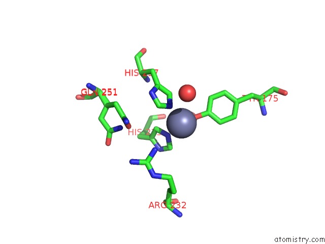
Mono view
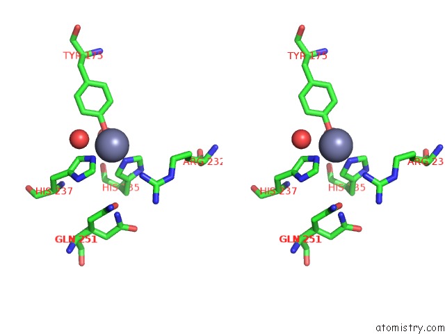
Stereo pair view
Mono view
Stereo pair view
A full contact list of Zinc with other atoms in the Zn binding
site number 1 of Crystal Structure of Catechol 1,2-Dioxygenase From Burkholderia Vietnamiensis within 5.0Å range:
|
Zinc binding site 2 out of 2 in 5td3
Go back to
Zinc binding site 2 out
of 2 in the Crystal Structure of Catechol 1,2-Dioxygenase From Burkholderia Vietnamiensis
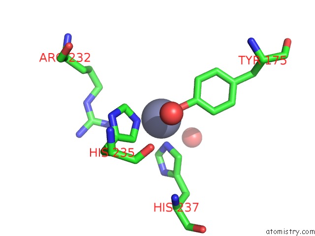
Mono view
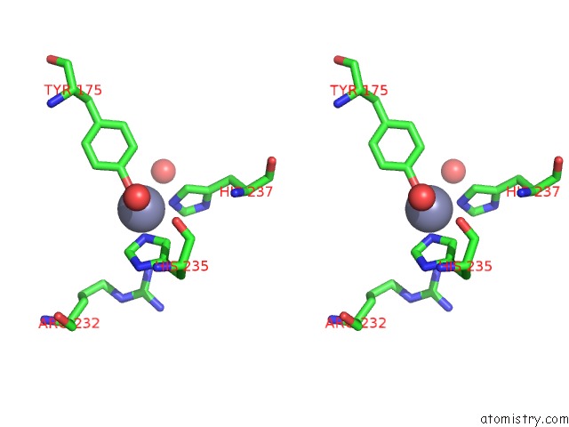
Stereo pair view
Mono view
Stereo pair view
A full contact list of Zinc with other atoms in the Zn binding
site number 2 of Crystal Structure of Catechol 1,2-Dioxygenase From Burkholderia Vietnamiensis within 5.0Å range:
|
Reference:
D.G.Conrady,
D.M.Dranow,
D.Lorimer,
T.E.Edwards.
Crystal Structure of Catechol 1,2-Dioxygenase From Burkholderia Vietnamiensis To Be Published.
Page generated: Mon Oct 28 08:26:08 2024
Last articles
I in 3WN5I in 3WYX
I in 3WGW
I in 3WD6
I in 3WB5
I in 3W31
I in 3WB4
I in 3W1N
I in 3W0F
I in 3W2Z