Zinc »
PDB 5sfw-5sh0 »
5sgf »
Zinc in PDB 5sgf: Crystal Structure of Human Phosphodiesterase 10 in Complex with Ethyl 1-Methyl-5-(Propylcarbamoyl)Pyrazole-4-Carboxylate
Enzymatic activity of Crystal Structure of Human Phosphodiesterase 10 in Complex with Ethyl 1-Methyl-5-(Propylcarbamoyl)Pyrazole-4-Carboxylate
All present enzymatic activity of Crystal Structure of Human Phosphodiesterase 10 in Complex with Ethyl 1-Methyl-5-(Propylcarbamoyl)Pyrazole-4-Carboxylate:
3.1.4.17;
3.1.4.17;
Protein crystallography data
The structure of Crystal Structure of Human Phosphodiesterase 10 in Complex with Ethyl 1-Methyl-5-(Propylcarbamoyl)Pyrazole-4-Carboxylate, PDB code: 5sgf
was solved by
C.Joseph,
J.Benz,
A.Flohr,
J.Peters,
M.G.Rudolph,
with X-Ray Crystallography technique. A brief refinement statistics is given in the table below:
| Resolution Low / High (Å) | 35.34 / 2.30 |
| Space group | H 3 |
| Cell size a, b, c (Å), α, β, γ (°) | 135.027, 135.027, 234.883, 90, 90, 120 |
| R / Rfree (%) | 19.9 / 25.7 |
Other elements in 5sgf:
The structure of Crystal Structure of Human Phosphodiesterase 10 in Complex with Ethyl 1-Methyl-5-(Propylcarbamoyl)Pyrazole-4-Carboxylate also contains other interesting chemical elements:
| Magnesium | (Mg) | 4 atoms |
Zinc Binding Sites:
The binding sites of Zinc atom in the Crystal Structure of Human Phosphodiesterase 10 in Complex with Ethyl 1-Methyl-5-(Propylcarbamoyl)Pyrazole-4-Carboxylate
(pdb code 5sgf). This binding sites where shown within
5.0 Angstroms radius around Zinc atom.
In total 4 binding sites of Zinc where determined in the Crystal Structure of Human Phosphodiesterase 10 in Complex with Ethyl 1-Methyl-5-(Propylcarbamoyl)Pyrazole-4-Carboxylate, PDB code: 5sgf:
Jump to Zinc binding site number: 1; 2; 3; 4;
In total 4 binding sites of Zinc where determined in the Crystal Structure of Human Phosphodiesterase 10 in Complex with Ethyl 1-Methyl-5-(Propylcarbamoyl)Pyrazole-4-Carboxylate, PDB code: 5sgf:
Jump to Zinc binding site number: 1; 2; 3; 4;
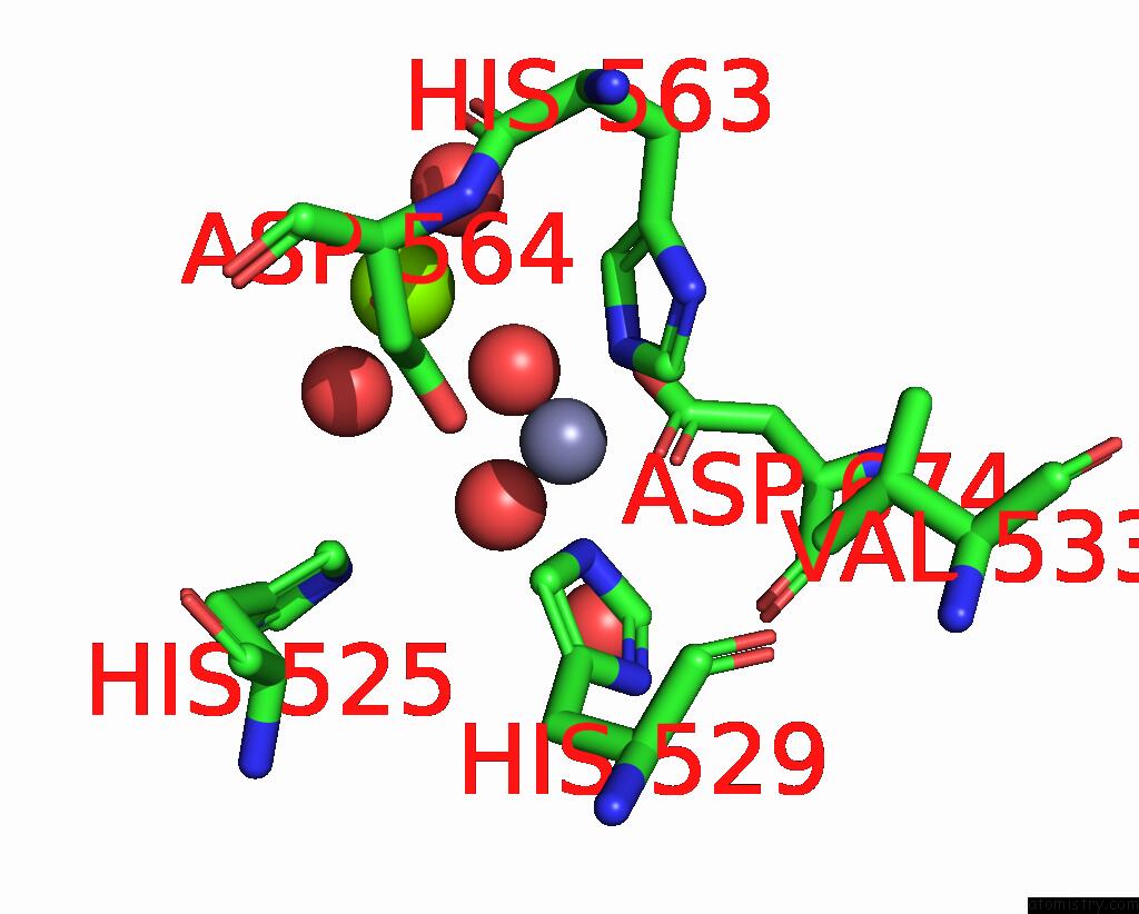
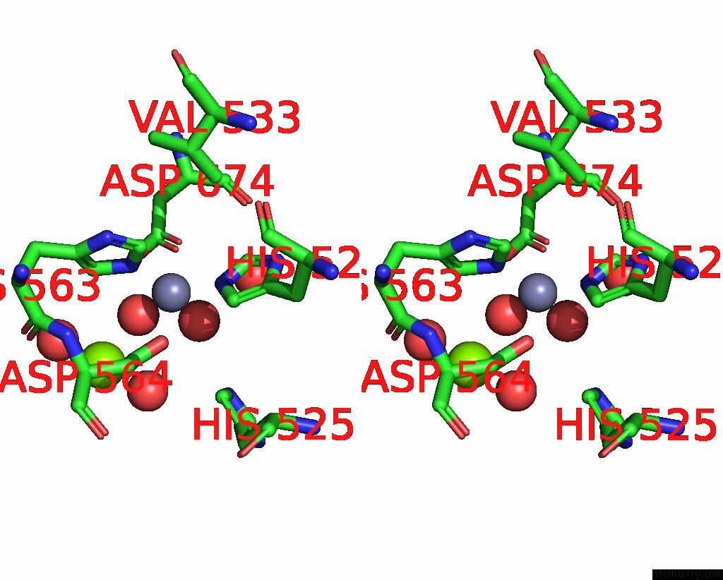
Zinc binding site 1 out of 4 in 5sgf
Go back to
Zinc binding site 1 out
of 4 in the Crystal Structure of Human Phosphodiesterase 10 in Complex with Ethyl 1-Methyl-5-(Propylcarbamoyl)Pyrazole-4-Carboxylate
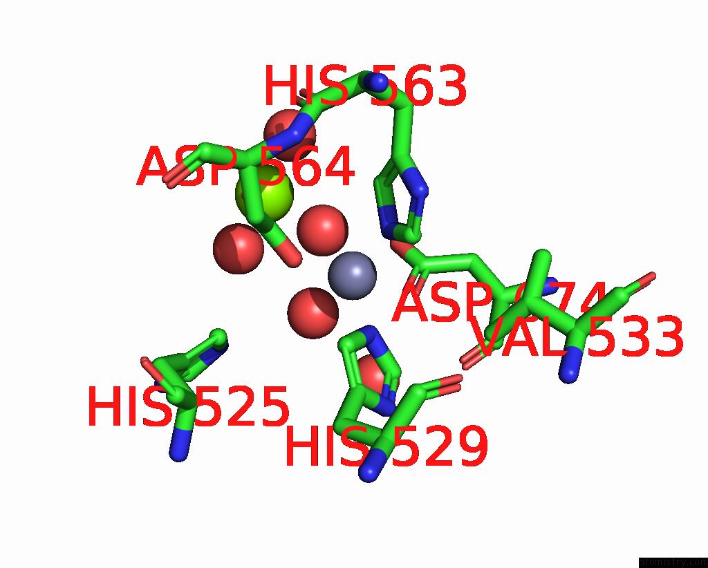
Mono view
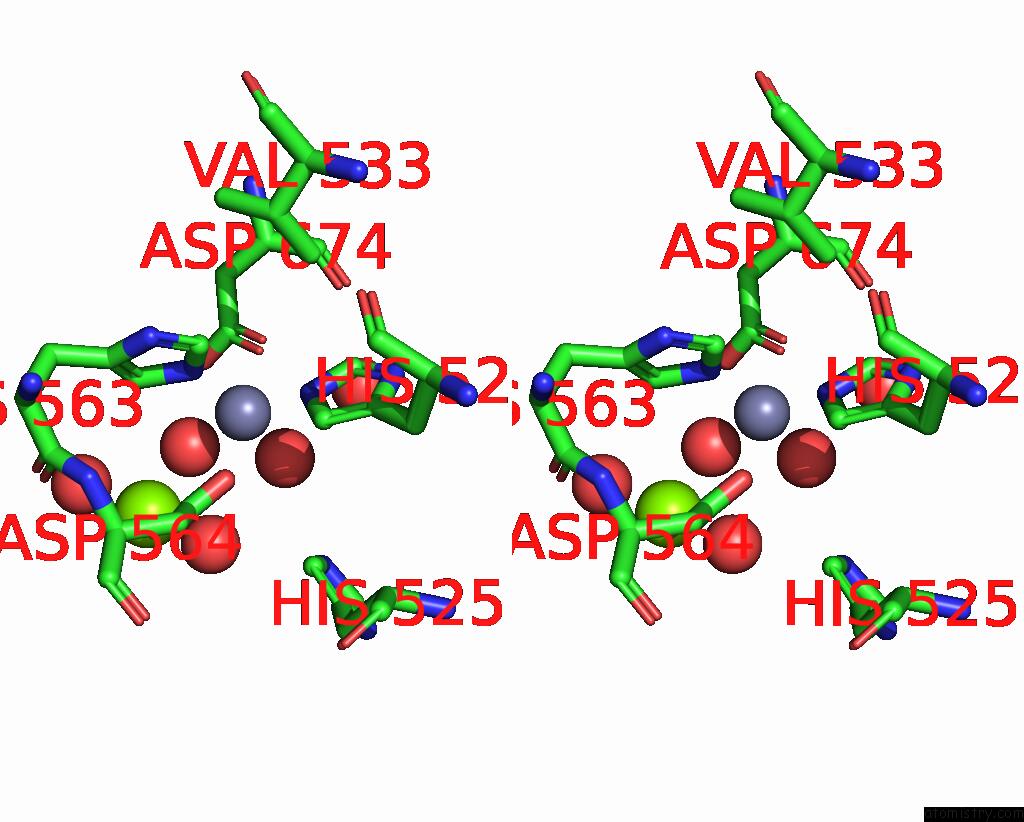
Stereo pair view
Mono view
Stereo pair view
A full contact list of Zinc with other atoms in the Zn binding
site number 1 of Crystal Structure of Human Phosphodiesterase 10 in Complex with Ethyl 1-Methyl-5-(Propylcarbamoyl)Pyrazole-4-Carboxylate within 5.0Å range:
|
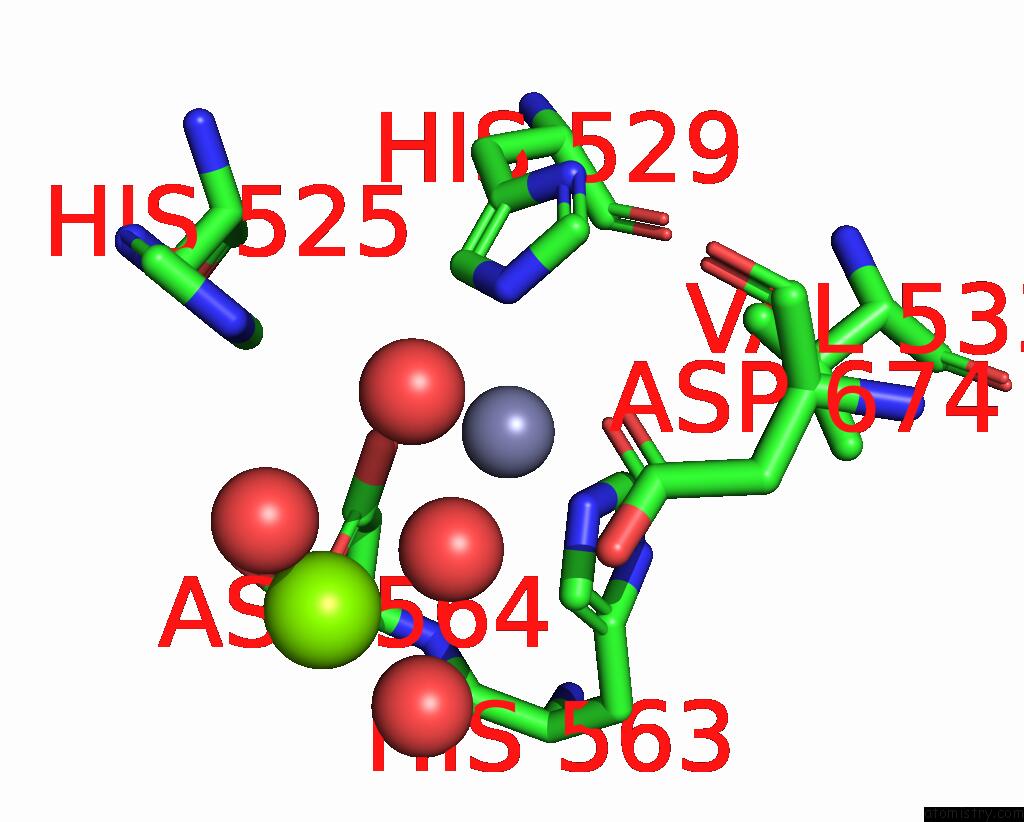
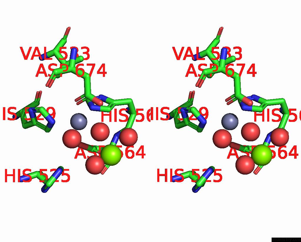
Zinc binding site 2 out of 4 in 5sgf
Go back to
Zinc binding site 2 out
of 4 in the Crystal Structure of Human Phosphodiesterase 10 in Complex with Ethyl 1-Methyl-5-(Propylcarbamoyl)Pyrazole-4-Carboxylate
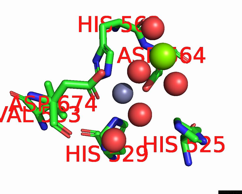
Mono view
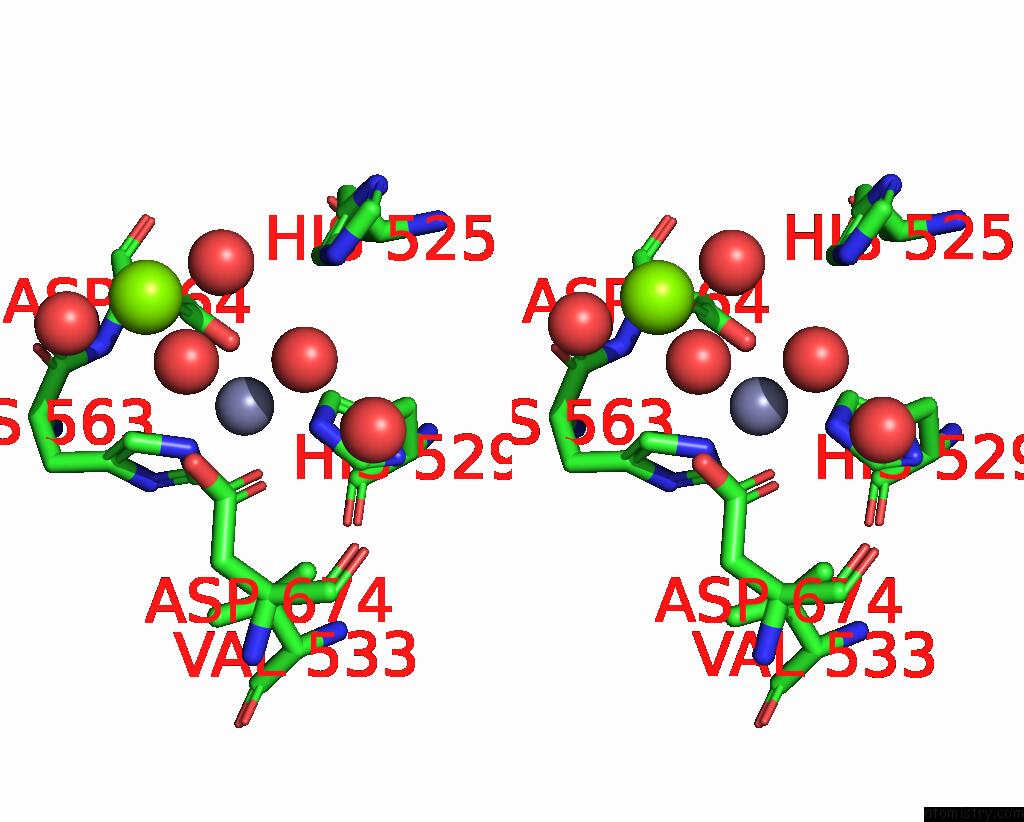
Stereo pair view
Mono view
Stereo pair view
A full contact list of Zinc with other atoms in the Zn binding
site number 2 of Crystal Structure of Human Phosphodiesterase 10 in Complex with Ethyl 1-Methyl-5-(Propylcarbamoyl)Pyrazole-4-Carboxylate within 5.0Å range:
|
Zinc binding site 3 out of 4 in 5sgf
Go back to
Zinc binding site 3 out
of 4 in the Crystal Structure of Human Phosphodiesterase 10 in Complex with Ethyl 1-Methyl-5-(Propylcarbamoyl)Pyrazole-4-Carboxylate
Mono view
Stereo pair view
Mono view
Stereo pair view
A full contact list of Zinc with other atoms in the Zn binding
site number 3 of Crystal Structure of Human Phosphodiesterase 10 in Complex with Ethyl 1-Methyl-5-(Propylcarbamoyl)Pyrazole-4-Carboxylate within 5.0Å range:
|
Zinc binding site 4 out of 4 in 5sgf
Go back to
Zinc binding site 4 out
of 4 in the Crystal Structure of Human Phosphodiesterase 10 in Complex with Ethyl 1-Methyl-5-(Propylcarbamoyl)Pyrazole-4-Carboxylate
Mono view
Stereo pair view
Mono view
Stereo pair view
A full contact list of Zinc with other atoms in the Zn binding
site number 4 of Crystal Structure of Human Phosphodiesterase 10 in Complex with Ethyl 1-Methyl-5-(Propylcarbamoyl)Pyrazole-4-Carboxylate within 5.0Å range:
|
Reference:
A.Flohr,
D.Schlatter,
B.Kuhn,
M.G.Rudolph.
Crystal Structure of A Human Phosphodiesterase 10 Complex To Be Published.
Page generated: Mon Oct 28 05:11:59 2024
Last articles
Mg in 7D1GMg in 7CWZ
Mg in 7CYQ
Mg in 7CWE
Mg in 7CWG
Mg in 7CWF
Mg in 7CWA
Mg in 7CVK
Mg in 7CVM
Mg in 7CVX