Zinc »
PDB 5n34-5nek »
5n6f »
Zinc in PDB 5n6f: Crystal Structure of Tgt in Complex with Guanine Fragment
Enzymatic activity of Crystal Structure of Tgt in Complex with Guanine Fragment
All present enzymatic activity of Crystal Structure of Tgt in Complex with Guanine Fragment:
2.4.2.29;
2.4.2.29;
Protein crystallography data
The structure of Crystal Structure of Tgt in Complex with Guanine Fragment, PDB code: 5n6f
was solved by
E.Hassaan,
A.Heine,
G.Klebe,
with X-Ray Crystallography technique. A brief refinement statistics is given in the table below:
| Resolution Low / High (Å) | 44.28 / 1.12 |
| Space group | C 1 2 1 |
| Cell size a, b, c (Å), α, β, γ (°) | 88.711, 64.851, 70.655, 90.00, 93.36, 90.00 |
| R / Rfree (%) | 13.6 / 15.3 |
Zinc Binding Sites:
The binding sites of Zinc atom in the Crystal Structure of Tgt in Complex with Guanine Fragment
(pdb code 5n6f). This binding sites where shown within
5.0 Angstroms radius around Zinc atom.
In total only one binding site of Zinc was determined in the Crystal Structure of Tgt in Complex with Guanine Fragment, PDB code: 5n6f:
In total only one binding site of Zinc was determined in the Crystal Structure of Tgt in Complex with Guanine Fragment, PDB code: 5n6f:
Zinc binding site 1 out of 1 in 5n6f
Go back to
Zinc binding site 1 out
of 1 in the Crystal Structure of Tgt in Complex with Guanine Fragment
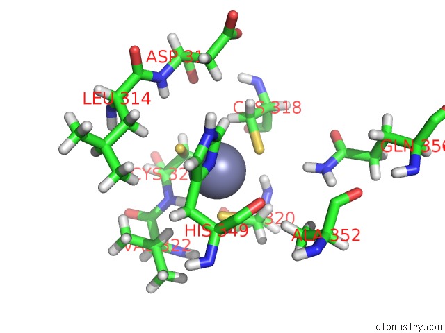
Mono view
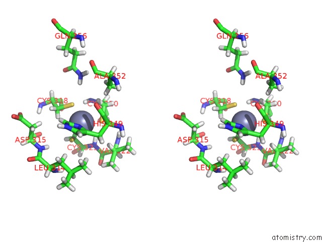
Stereo pair view
Mono view
Stereo pair view
A full contact list of Zinc with other atoms in the Zn binding
site number 1 of Crystal Structure of Tgt in Complex with Guanine Fragment within 5.0Å range:
|
Reference:
E.Hassaan,
A.Heine,
G.Klebe.
Crystal Structure of Tgt in Complex with Guanine Fragment To Be Published.
Page generated: Sun Oct 27 22:37:26 2024
Last articles
Mg in 9F37Mg in 9EZL
Mg in 9F28
Mg in 9F2R
Mg in 9F12
Mg in 9F11
Mg in 9F10
Mg in 9F0Z
Mg in 9F0Y
Mg in 9F07