Zinc »
PDB 5mt3-5n31 »
5mvw »
Zinc in PDB 5mvw: Complex Between the Leucine Zipper (Lz) and Centrosomin-Motif 2 (CM2) Domains of Drosophila Melanogaster Centrosomin (Cnn)
Protein crystallography data
The structure of Complex Between the Leucine Zipper (Lz) and Centrosomin-Motif 2 (CM2) Domains of Drosophila Melanogaster Centrosomin (Cnn), PDB code: 5mvw
was solved by
Z.Feng,
S.Johnson,
J.W.Raff,
S.M.Lea,
with X-Ray Crystallography technique. A brief refinement statistics is given in the table below:
| Resolution Low / High (Å) | 42.74 / 1.82 |
| Space group | P 1 21 1 |
| Cell size a, b, c (Å), α, β, γ (°) | 44.880, 44.270, 72.530, 90.00, 107.77, 90.00 |
| R / Rfree (%) | 22.2 / 24.6 |
Other elements in 5mvw:
The structure of Complex Between the Leucine Zipper (Lz) and Centrosomin-Motif 2 (CM2) Domains of Drosophila Melanogaster Centrosomin (Cnn) also contains other interesting chemical elements:
| Chlorine | (Cl) | 1 atom |
Zinc Binding Sites:
The binding sites of Zinc atom in the Complex Between the Leucine Zipper (Lz) and Centrosomin-Motif 2 (CM2) Domains of Drosophila Melanogaster Centrosomin (Cnn)
(pdb code 5mvw). This binding sites where shown within
5.0 Angstroms radius around Zinc atom.
In total only one binding site of Zinc was determined in the Complex Between the Leucine Zipper (Lz) and Centrosomin-Motif 2 (CM2) Domains of Drosophila Melanogaster Centrosomin (Cnn), PDB code: 5mvw:
In total only one binding site of Zinc was determined in the Complex Between the Leucine Zipper (Lz) and Centrosomin-Motif 2 (CM2) Domains of Drosophila Melanogaster Centrosomin (Cnn), PDB code: 5mvw:
Zinc binding site 1 out of 1 in 5mvw
Go back to
Zinc binding site 1 out
of 1 in the Complex Between the Leucine Zipper (Lz) and Centrosomin-Motif 2 (CM2) Domains of Drosophila Melanogaster Centrosomin (Cnn)
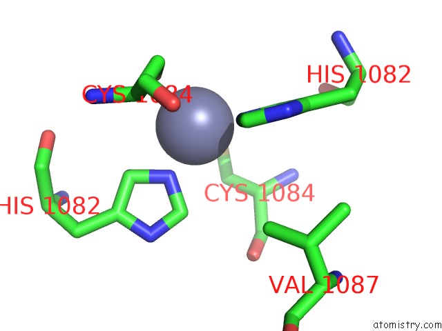
Mono view
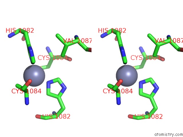
Stereo pair view
Mono view
Stereo pair view
A full contact list of Zinc with other atoms in the Zn binding
site number 1 of Complex Between the Leucine Zipper (Lz) and Centrosomin-Motif 2 (CM2) Domains of Drosophila Melanogaster Centrosomin (Cnn) within 5.0Å range:
|
Reference:
Z.Feng,
A.Caballe,
A.Wainman,
S.Johnson,
A.F.M.Haensele,
M.A.Cottee,
P.T.Conduit,
S.M.Lea,
J.W.Raff.
Structural Basis For Mitotic Centrosome Assembly in Flies. Cell V. 169 1078 2017.
ISSN: ISSN 1097-4172
PubMed: 28575671
DOI: 10.1016/J.CELL.2017.05.030
Page generated: Sun Oct 27 22:21:50 2024
ISSN: ISSN 1097-4172
PubMed: 28575671
DOI: 10.1016/J.CELL.2017.05.030
Last articles
Mg in 4KP6Mg in 4KO8
Mg in 4KP4
Mg in 4KP1
Mg in 4KHP
Mg in 4KNW
Mg in 4KNX
Mg in 4KNV
Mg in 4KNR
Mg in 4KMQ