Zinc »
PDB 5mt3-5n31 »
5mte »
Zinc in PDB 5mte: Crystal Structure of Pdf From the Vibrio Parahaemolyticus Bacteriophage VP16T in Complex with Actinonin - Crystal Form II
Protein crystallography data
The structure of Crystal Structure of Pdf From the Vibrio Parahaemolyticus Bacteriophage VP16T in Complex with Actinonin - Crystal Form II, PDB code: 5mte
was solved by
S.Fieulaine,
R.Grzela,
C.Giglione,
T.Meinnel,
with X-Ray Crystallography technique. A brief refinement statistics is given in the table below:
| Resolution Low / High (Å) | 41.67 / 1.40 |
| Space group | P 32 2 1 |
| Cell size a, b, c (Å), α, β, γ (°) | 64.620, 64.620, 124.890, 90.00, 90.00, 120.00 |
| R / Rfree (%) | 18.7 / 20.3 |
Other elements in 5mte:
The structure of Crystal Structure of Pdf From the Vibrio Parahaemolyticus Bacteriophage VP16T in Complex with Actinonin - Crystal Form II also contains other interesting chemical elements:
| Nickel | (Ni) | 3 atoms |
Zinc Binding Sites:
The binding sites of Zinc atom in the Crystal Structure of Pdf From the Vibrio Parahaemolyticus Bacteriophage VP16T in Complex with Actinonin - Crystal Form II
(pdb code 5mte). This binding sites where shown within
5.0 Angstroms radius around Zinc atom.
In total 2 binding sites of Zinc where determined in the Crystal Structure of Pdf From the Vibrio Parahaemolyticus Bacteriophage VP16T in Complex with Actinonin - Crystal Form II, PDB code: 5mte:
Jump to Zinc binding site number: 1; 2;
In total 2 binding sites of Zinc where determined in the Crystal Structure of Pdf From the Vibrio Parahaemolyticus Bacteriophage VP16T in Complex with Actinonin - Crystal Form II, PDB code: 5mte:
Jump to Zinc binding site number: 1; 2;
Zinc binding site 1 out of 2 in 5mte
Go back to
Zinc binding site 1 out
of 2 in the Crystal Structure of Pdf From the Vibrio Parahaemolyticus Bacteriophage VP16T in Complex with Actinonin - Crystal Form II
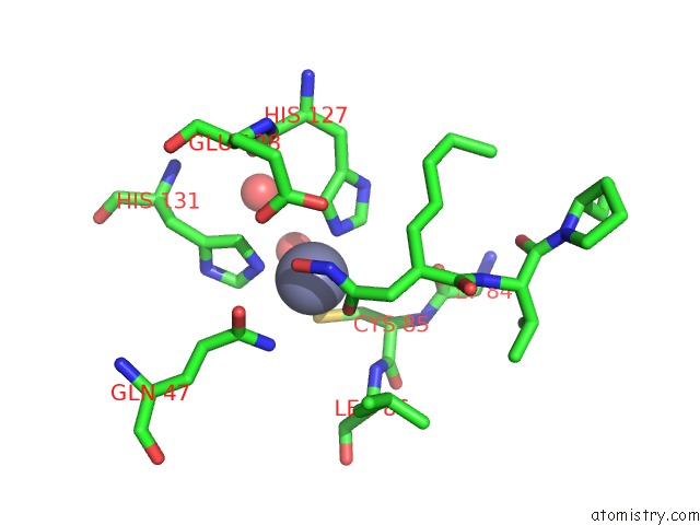
Mono view
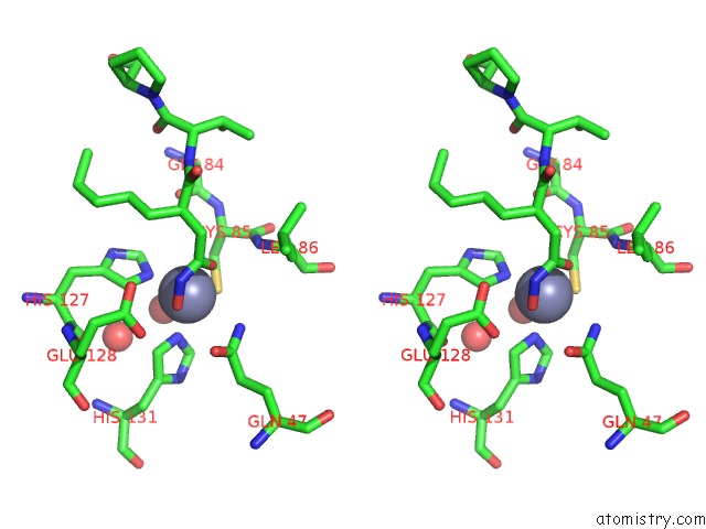
Stereo pair view
Mono view
Stereo pair view
A full contact list of Zinc with other atoms in the Zn binding
site number 1 of Crystal Structure of Pdf From the Vibrio Parahaemolyticus Bacteriophage VP16T in Complex with Actinonin - Crystal Form II within 5.0Å range:
|
Zinc binding site 2 out of 2 in 5mte
Go back to
Zinc binding site 2 out
of 2 in the Crystal Structure of Pdf From the Vibrio Parahaemolyticus Bacteriophage VP16T in Complex with Actinonin - Crystal Form II
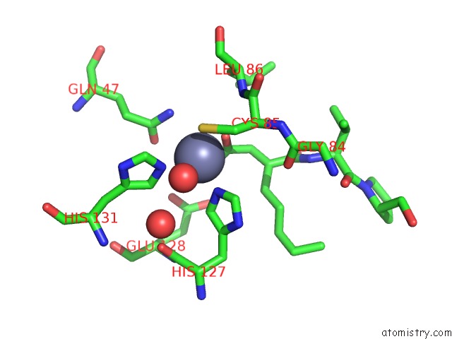
Mono view
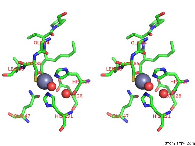
Stereo pair view
Mono view
Stereo pair view
A full contact list of Zinc with other atoms in the Zn binding
site number 2 of Crystal Structure of Pdf From the Vibrio Parahaemolyticus Bacteriophage VP16T in Complex with Actinonin - Crystal Form II within 5.0Å range:
|
Reference:
R.Grzela,
J.Nusbaum,
S.Fieulaine,
F.Lavecchia,
M.Desmadril,
N.Nhiri,
A.Van Dorsselaer,
S.Cianferani,
E.Jacquet,
T.Meinnel,
C.Giglione.
Peptide Deformylases From Vibrio Parahaemolyticus Phage and Bacteria Display Similar Deformylase Activity and Inhibitor Binding Clefts. Biochim. Biophys. Acta V.1866 348 2018.
ISSN: ISSN 0006-3002
PubMed: 29101077
DOI: 10.1016/J.BBAPAP.2017.10.007
Page generated: Sun Oct 27 22:20:12 2024
ISSN: ISSN 0006-3002
PubMed: 29101077
DOI: 10.1016/J.BBAPAP.2017.10.007
Last articles
F in 6UKWF in 6UL5
F in 6UJJ
F in 6UJX
F in 6UIL
F in 6UJZ
F in 6UGZ
F in 6UI2
F in 6UIR
F in 6UF0