Zinc »
PDB 5kb0-5kj1 »
5kb1 »
Zinc in PDB 5kb1: Crystal Structure of A Tris-Thiolate Hg(II) Complex in A De Novo Three Stranded Coiled Coil Peptide
Protein crystallography data
The structure of Crystal Structure of A Tris-Thiolate Hg(II) Complex in A De Novo Three Stranded Coiled Coil Peptide, PDB code: 5kb1
was solved by
L.Ruckcthong,
M.L.Zastrow,
J.A.Stuckey,
V.L.Pecoraro,
with X-Ray Crystallography technique. A brief refinement statistics is given in the table below:
| Resolution Low / High (Å) | 47.88 / 2.09 |
| Space group | H 3 2 |
| Cell size a, b, c (Å), α, β, γ (°) | 38.048, 38.048, 143.651, 90.00, 90.00, 120.00 |
| R / Rfree (%) | 21.8 / 25.7 |
Other elements in 5kb1:
The structure of Crystal Structure of A Tris-Thiolate Hg(II) Complex in A De Novo Three Stranded Coiled Coil Peptide also contains other interesting chemical elements:
| Mercury | (Hg) | 1 atom |
| Chlorine | (Cl) | 1 atom |
Zinc Binding Sites:
The binding sites of Zinc atom in the Crystal Structure of A Tris-Thiolate Hg(II) Complex in A De Novo Three Stranded Coiled Coil Peptide
(pdb code 5kb1). This binding sites where shown within
5.0 Angstroms radius around Zinc atom.
In total 2 binding sites of Zinc where determined in the Crystal Structure of A Tris-Thiolate Hg(II) Complex in A De Novo Three Stranded Coiled Coil Peptide, PDB code: 5kb1:
Jump to Zinc binding site number: 1; 2;
In total 2 binding sites of Zinc where determined in the Crystal Structure of A Tris-Thiolate Hg(II) Complex in A De Novo Three Stranded Coiled Coil Peptide, PDB code: 5kb1:
Jump to Zinc binding site number: 1; 2;
Zinc binding site 1 out of 2 in 5kb1
Go back to
Zinc binding site 1 out
of 2 in the Crystal Structure of A Tris-Thiolate Hg(II) Complex in A De Novo Three Stranded Coiled Coil Peptide
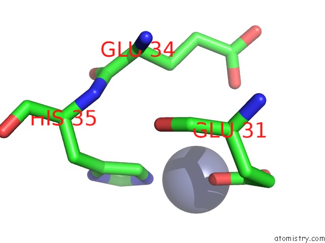
Mono view
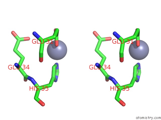
Stereo pair view
Mono view
Stereo pair view
A full contact list of Zinc with other atoms in the Zn binding
site number 1 of Crystal Structure of A Tris-Thiolate Hg(II) Complex in A De Novo Three Stranded Coiled Coil Peptide within 5.0Å range:
|
Zinc binding site 2 out of 2 in 5kb1
Go back to
Zinc binding site 2 out
of 2 in the Crystal Structure of A Tris-Thiolate Hg(II) Complex in A De Novo Three Stranded Coiled Coil Peptide
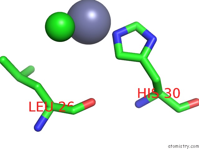
Mono view
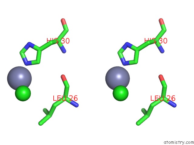
Stereo pair view
Mono view
Stereo pair view
A full contact list of Zinc with other atoms in the Zn binding
site number 2 of Crystal Structure of A Tris-Thiolate Hg(II) Complex in A De Novo Three Stranded Coiled Coil Peptide within 5.0Å range:
|
Reference:
L.Ruckthong,
M.L.Zastrow,
J.A.Stuckey,
V.L.Pecoraro.
A Crystallographic Examination of Predisposition Versus Preorganization in De Novo Designed Metalloproteins. J.Am.Chem.Soc. V. 138 11979 2016.
ISSN: ESSN 1520-5126
PubMed: 27532255
DOI: 10.1021/JACS.6B07165
Page generated: Sun Oct 27 20:15:29 2024
ISSN: ESSN 1520-5126
PubMed: 27532255
DOI: 10.1021/JACS.6B07165
Last articles
Fe in 2YXOFe in 2YRS
Fe in 2YXC
Fe in 2YNM
Fe in 2YVJ
Fe in 2YP1
Fe in 2YU2
Fe in 2YU1
Fe in 2YQB
Fe in 2YOO