Zinc »
PDB 5jn7-5k1b »
5k16 »
Zinc in PDB 5k16: Crystal Structure of Free Ubiquitin-Specific Protease 12
Enzymatic activity of Crystal Structure of Free Ubiquitin-Specific Protease 12
All present enzymatic activity of Crystal Structure of Free Ubiquitin-Specific Protease 12:
3.4.19.12;
3.4.19.12;
Protein crystallography data
The structure of Crystal Structure of Free Ubiquitin-Specific Protease 12, PDB code: 5k16
was solved by
H.Li,
A.D.D'andrea,
N.Zheng,
with X-Ray Crystallography technique. A brief refinement statistics is given in the table below:
| Resolution Low / High (Å) | 42.45 / 2.60 |
| Space group | P 21 21 21 |
| Cell size a, b, c (Å), α, β, γ (°) | 52.396, 109.636, 134.193, 90.00, 90.00, 90.00 |
| R / Rfree (%) | 19 / 24.6 |
Zinc Binding Sites:
The binding sites of Zinc atom in the Crystal Structure of Free Ubiquitin-Specific Protease 12
(pdb code 5k16). This binding sites where shown within
5.0 Angstroms radius around Zinc atom.
In total 2 binding sites of Zinc where determined in the Crystal Structure of Free Ubiquitin-Specific Protease 12, PDB code: 5k16:
Jump to Zinc binding site number: 1; 2;
In total 2 binding sites of Zinc where determined in the Crystal Structure of Free Ubiquitin-Specific Protease 12, PDB code: 5k16:
Jump to Zinc binding site number: 1; 2;
Zinc binding site 1 out of 2 in 5k16
Go back to
Zinc binding site 1 out
of 2 in the Crystal Structure of Free Ubiquitin-Specific Protease 12
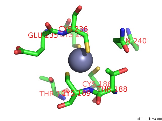
Mono view
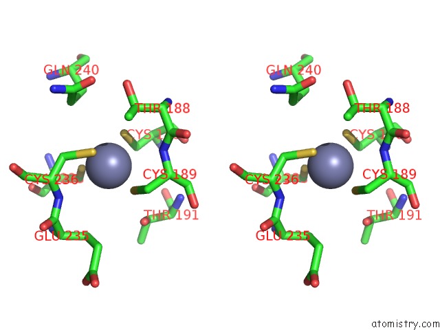
Stereo pair view
Mono view
Stereo pair view
A full contact list of Zinc with other atoms in the Zn binding
site number 1 of Crystal Structure of Free Ubiquitin-Specific Protease 12 within 5.0Å range:
|
Zinc binding site 2 out of 2 in 5k16
Go back to
Zinc binding site 2 out
of 2 in the Crystal Structure of Free Ubiquitin-Specific Protease 12
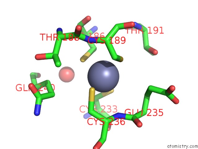
Mono view
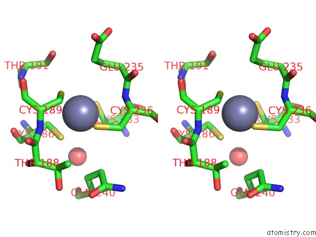
Stereo pair view
Mono view
Stereo pair view
A full contact list of Zinc with other atoms in the Zn binding
site number 2 of Crystal Structure of Free Ubiquitin-Specific Protease 12 within 5.0Å range:
|
Reference:
H.Li,
K.S.Lim,
H.Kim,
T.R.Hinds,
U.Jo,
H.Mao,
C.E.Weller,
J.Sun,
C.Chatterjee,
A.D.D'andrea,
N.Zheng.
Allosteric Activation of Ubiquitin-Specific Proteases By Beta-Propeller Proteins UAF1 and WDR20. Mol.Cell V. 63 249 2016.
ISSN: ISSN 1097-2765
PubMed: 27373336
DOI: 10.1016/J.MOLCEL.2016.05.031
Page generated: Thu Aug 21 04:02:59 2025
ISSN: ISSN 1097-2765
PubMed: 27373336
DOI: 10.1016/J.MOLCEL.2016.05.031
Last articles
Zn in 5SIKZn in 5SIJ
Zn in 5SIM
Zn in 5SIL
Zn in 5SIH
Zn in 5SII
Zn in 5SIG
Zn in 5SIF
Zn in 5SID
Zn in 5SIE