Zinc »
PDB 5gmk-5h7s »
5h7r »
Zinc in PDB 5h7r: Structural Basis of the Flanking Zinc-Finger Motifs Crucial For the E3 Ligase Activity of the LNX1 Ring Domain
Protein crystallography data
The structure of Structural Basis of the Flanking Zinc-Finger Motifs Crucial For the E3 Ligase Activity of the LNX1 Ring Domain, PDB code: 5h7r
was solved by
D.Nayak,
J.Sivaraman,
with X-Ray Crystallography technique. A brief refinement statistics is given in the table below:
| Resolution Low / High (Å) | 79.88 / 1.70 |
| Space group | P 62 2 2 |
| Cell size a, b, c (Å), α, β, γ (°) | 92.375, 92.375, 84.410, 90.00, 90.00, 120.00 |
| R / Rfree (%) | 17.8 / 21.5 |
Zinc Binding Sites:
The binding sites of Zinc atom in the Structural Basis of the Flanking Zinc-Finger Motifs Crucial For the E3 Ligase Activity of the LNX1 Ring Domain
(pdb code 5h7r). This binding sites where shown within
5.0 Angstroms radius around Zinc atom.
In total 4 binding sites of Zinc where determined in the Structural Basis of the Flanking Zinc-Finger Motifs Crucial For the E3 Ligase Activity of the LNX1 Ring Domain, PDB code: 5h7r:
Jump to Zinc binding site number: 1; 2; 3; 4;
In total 4 binding sites of Zinc where determined in the Structural Basis of the Flanking Zinc-Finger Motifs Crucial For the E3 Ligase Activity of the LNX1 Ring Domain, PDB code: 5h7r:
Jump to Zinc binding site number: 1; 2; 3; 4;
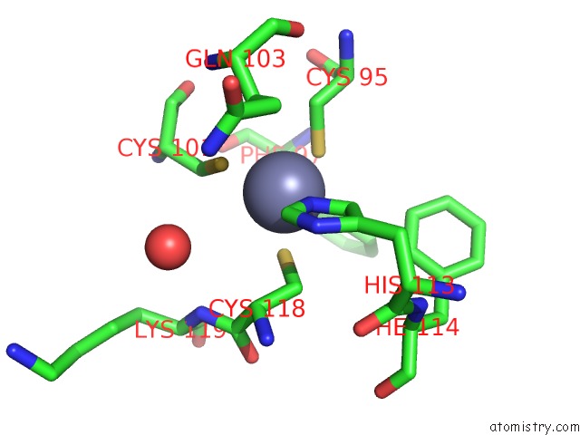
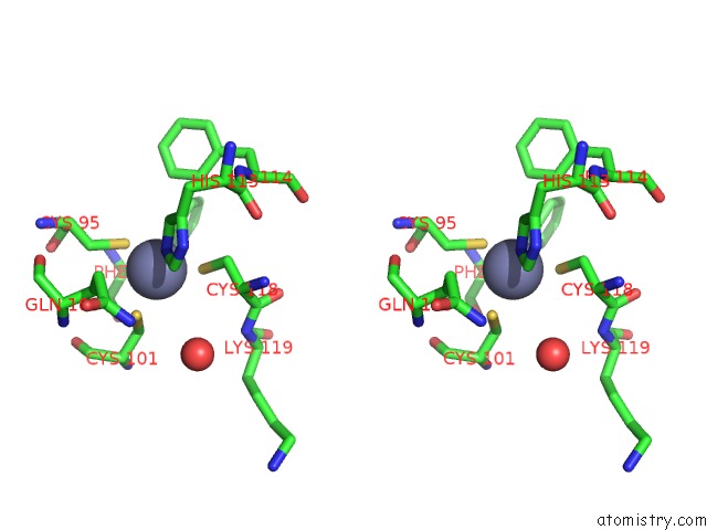
Zinc binding site 1 out of 4 in 5h7r
Go back to
Zinc binding site 1 out
of 4 in the Structural Basis of the Flanking Zinc-Finger Motifs Crucial For the E3 Ligase Activity of the LNX1 Ring Domain
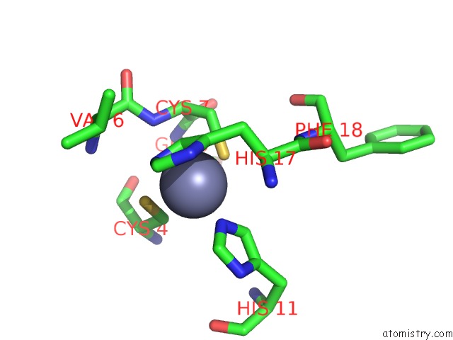
Mono view
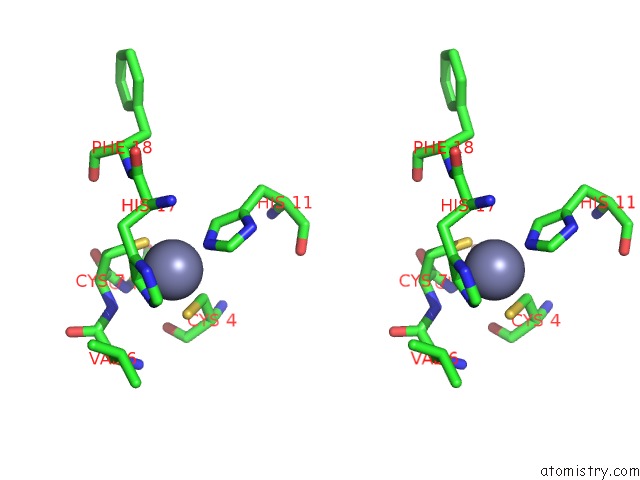
Stereo pair view
Mono view
Stereo pair view
A full contact list of Zinc with other atoms in the Zn binding
site number 1 of Structural Basis of the Flanking Zinc-Finger Motifs Crucial For the E3 Ligase Activity of the LNX1 Ring Domain within 5.0Å range:
|
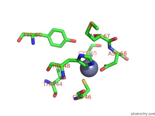
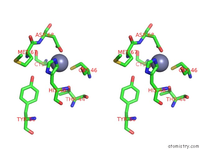
Zinc binding site 2 out of 4 in 5h7r
Go back to
Zinc binding site 2 out
of 4 in the Structural Basis of the Flanking Zinc-Finger Motifs Crucial For the E3 Ligase Activity of the LNX1 Ring Domain
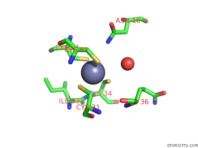
Mono view
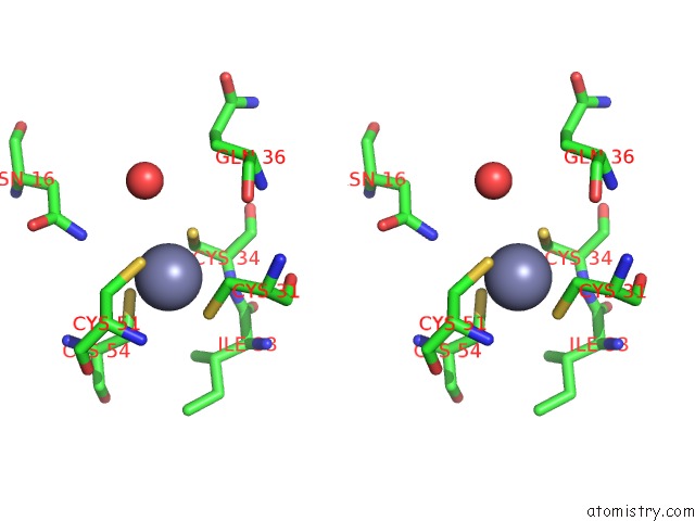
Stereo pair view
Mono view
Stereo pair view
A full contact list of Zinc with other atoms in the Zn binding
site number 2 of Structural Basis of the Flanking Zinc-Finger Motifs Crucial For the E3 Ligase Activity of the LNX1 Ring Domain within 5.0Å range:
|
Zinc binding site 3 out of 4 in 5h7r
Go back to
Zinc binding site 3 out
of 4 in the Structural Basis of the Flanking Zinc-Finger Motifs Crucial For the E3 Ligase Activity of the LNX1 Ring Domain
Mono view
Stereo pair view
Mono view
Stereo pair view
A full contact list of Zinc with other atoms in the Zn binding
site number 3 of Structural Basis of the Flanking Zinc-Finger Motifs Crucial For the E3 Ligase Activity of the LNX1 Ring Domain within 5.0Å range:
|
Zinc binding site 4 out of 4 in 5h7r
Go back to
Zinc binding site 4 out
of 4 in the Structural Basis of the Flanking Zinc-Finger Motifs Crucial For the E3 Ligase Activity of the LNX1 Ring Domain
Mono view
Stereo pair view
Mono view
Stereo pair view
A full contact list of Zinc with other atoms in the Zn binding
site number 4 of Structural Basis of the Flanking Zinc-Finger Motifs Crucial For the E3 Ligase Activity of the LNX1 Ring Domain within 5.0Å range:
|
Reference:
D.Nayak,
J.Sivaraman.
Structure of LNX1:UBC13~Ubiquitin Complex Reveals the Role of Additional Motifs For the E3 Ligase Activity of LNX1. J. Mol. Biol. V. 430 1173 2018.
ISSN: ESSN 1089-8638
PubMed: 29496391
DOI: 10.1016/J.JMB.2018.02.016
Page generated: Thu Aug 21 03:01:54 2025
ISSN: ESSN 1089-8638
PubMed: 29496391
DOI: 10.1016/J.JMB.2018.02.016
Last articles
Zn in 5SVIZn in 5SVY
Zn in 5SVE
Zn in 5SVX
Zn in 5SVA
Zn in 5SVB
Zn in 5SVC
Zn in 5SMK
Zn in 5SMI
Zn in 5SMH