Zinc »
PDB 5gmk-5h7s »
5gox »
Zinc in PDB 5gox: Eukaryotic RAD50 Functions As A Rod-Shaped Dimer
Protein crystallography data
The structure of Eukaryotic RAD50 Functions As A Rod-Shaped Dimer, PDB code: 5gox
was solved by
Y.B.Park,
M.Hohl,
M.Padjasek,
E.Jeong,
K.S.Jin,
A.Krezel,
J.H.J.Petrini,
Y.Cho,
with X-Ray Crystallography technique. A brief refinement statistics is given in the table below:
| Resolution Low / High (Å) | 28.22 / 2.41 |
| Space group | P 1 21 1 |
| Cell size a, b, c (Å), α, β, γ (°) | 42.180, 61.972, 81.540, 90.00, 99.75, 90.00 |
| R / Rfree (%) | 21.3 / 27.1 |
Zinc Binding Sites:
The binding sites of Zinc atom in the Eukaryotic RAD50 Functions As A Rod-Shaped Dimer
(pdb code 5gox). This binding sites where shown within
5.0 Angstroms radius around Zinc atom.
In total only one binding site of Zinc was determined in the Eukaryotic RAD50 Functions As A Rod-Shaped Dimer, PDB code: 5gox:
In total only one binding site of Zinc was determined in the Eukaryotic RAD50 Functions As A Rod-Shaped Dimer, PDB code: 5gox:
Zinc binding site 1 out of 1 in 5gox
Go back to
Zinc binding site 1 out
of 1 in the Eukaryotic RAD50 Functions As A Rod-Shaped Dimer
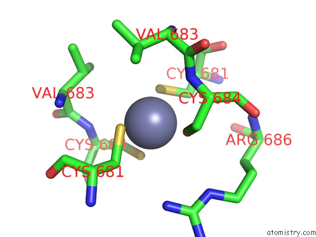
Mono view
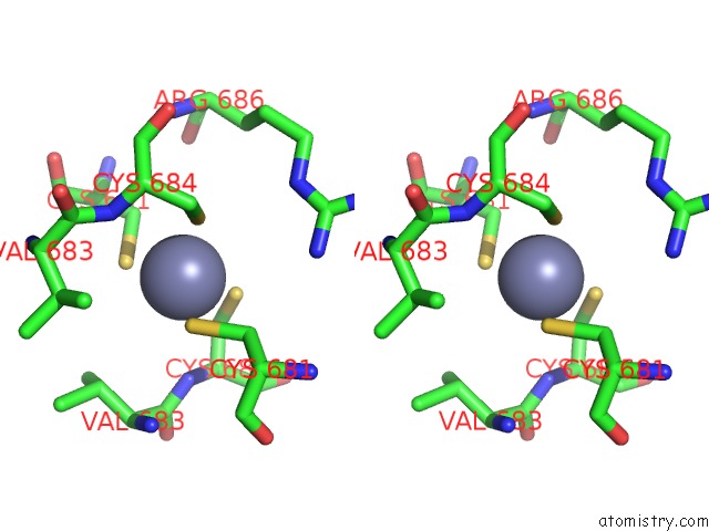
Stereo pair view
Mono view
Stereo pair view
A full contact list of Zinc with other atoms in the Zn binding
site number 1 of Eukaryotic RAD50 Functions As A Rod-Shaped Dimer within 5.0Å range:
|
Reference:
Y.B.Park,
M.Hohl,
M.Padjasek,
E.Jeong,
K.S.Jin,
A.Krezel,
J.H.J.Petrini,
Y.Cho.
Eukaryotic RAD50 Functions As A Rod-Shaped Dimer Nat. Struct. Mol. Biol. V. 24 248 2017.
ISSN: ESSN 1545-9985
PubMed: 28134932
DOI: 10.1038/NSMB.3369
Page generated: Thu Aug 21 02:55:28 2025
ISSN: ESSN 1545-9985
PubMed: 28134932
DOI: 10.1038/NSMB.3369
Last articles
Zn in 5SKWZn in 5SKV
Zn in 5SKU
Zn in 5SKT
Zn in 5SKS
Zn in 5SKR
Zn in 5SKQ
Zn in 5SKP
Zn in 5SKN
Zn in 5SKO