Zinc »
PDB 5eht-5ew0 »
5esb »
Zinc in PDB 5esb: Crystal Structure of A Genotype 1A/3A Chimeric Hcv NS3/4A Protease in Complex with Vaniprevir
Protein crystallography data
The structure of Crystal Structure of A Genotype 1A/3A Chimeric Hcv NS3/4A Protease in Complex with Vaniprevir, PDB code: 5esb
was solved by
D.Soumana,
N.K.Yilmaz,
A.Ali,
K.L.Prachanronarong,
C.A.Schiffer,
with X-Ray Crystallography technique. A brief refinement statistics is given in the table below:
| Resolution Low / High (Å) | 33.00 / 2.40 |
| Space group | P 21 21 21 |
| Cell size a, b, c (Å), α, β, γ (°) | 55.119, 58.403, 60.016, 90.00, 90.00, 90.00 |
| R / Rfree (%) | 18.2 / 23.3 |
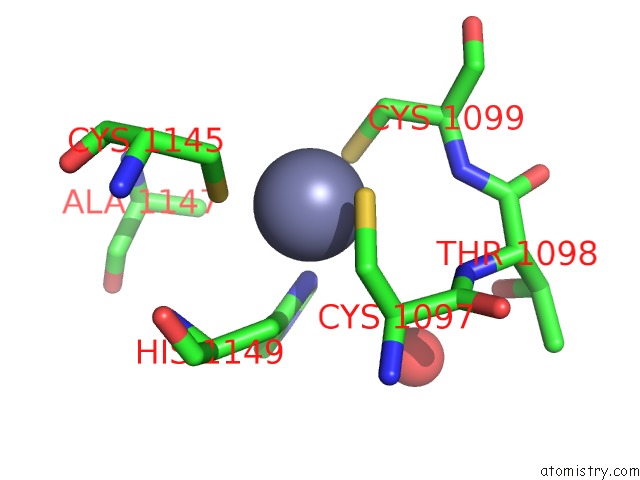
Zinc Binding Sites:
The binding sites of Zinc atom in the Crystal Structure of A Genotype 1A/3A Chimeric Hcv NS3/4A Protease in Complex with Vaniprevir
(pdb code 5esb). This binding sites where shown within
5.0 Angstroms radius around Zinc atom.
In total only one binding site of Zinc was determined in the Crystal Structure of A Genotype 1A/3A Chimeric Hcv NS3/4A Protease in Complex with Vaniprevir, PDB code: 5esb:
In total only one binding site of Zinc was determined in the Crystal Structure of A Genotype 1A/3A Chimeric Hcv NS3/4A Protease in Complex with Vaniprevir, PDB code: 5esb:
Zinc binding site 1 out of 1 in 5esb
Go back to
Zinc binding site 1 out
of 1 in the Crystal Structure of A Genotype 1A/3A Chimeric Hcv NS3/4A Protease in Complex with Vaniprevir
Mono view

Stereo pair view
Mono view
Stereo pair view
A full contact list of Zinc with other atoms in the Zn binding
site number 1 of Crystal Structure of A Genotype 1A/3A Chimeric Hcv NS3/4A Protease in Complex with Vaniprevir within 5.0Å range:
|
Reference:
D.I.Soumana,
N.Kurt Yilmaz,
A.Ali,
K.L.Prachanronarong,
C.A.Schiffer.
Molecular and Dynamic Mechanism Underlying Drug Resistance in Genotype 3 Hepatitis C NS3/4A Protease. J.Am.Chem.Soc. V. 138 11850 2016.
ISSN: ESSN 1520-5126
PubMed: 27512818
DOI: 10.1021/JACS.6B06454
Page generated: Sun Oct 27 15:29:39 2024
ISSN: ESSN 1520-5126
PubMed: 27512818
DOI: 10.1021/JACS.6B06454
Last articles
Fe in 7LSNFe in 7LSL
Fe in 7LSJ
Fe in 7LS4
Fe in 7LS3
Fe in 7LRV
Fe in 7LRR
Fe in 7LRL
Fe in 7LRB
Fe in 7LRA