Zinc »
PDB 5eht-5ew0 »
5ejk »
Zinc in PDB 5ejk: Crystal Structure of the Rous Sarcoma Virus Intasome
Enzymatic activity of Crystal Structure of the Rous Sarcoma Virus Intasome
All present enzymatic activity of Crystal Structure of the Rous Sarcoma Virus Intasome:
2.7.7.49; 2.7.7.7; 3.1.26.4;
2.7.7.49; 2.7.7.7; 3.1.26.4;
Protein crystallography data
The structure of Crystal Structure of the Rous Sarcoma Virus Intasome, PDB code: 5ejk
was solved by
Z.Yin,
K.Shi,
S.Banerjee,
H.Aihara,
with X-Ray Crystallography technique. A brief refinement statistics is given in the table below:
| Resolution Low / High (Å) | 49.22 / 3.80 |
| Space group | P 1 21 1 |
| Cell size a, b, c (Å), α, β, γ (°) | 124.936, 157.854, 126.583, 90.00, 110.94, 90.00 |
| R / Rfree (%) | 25.4 / 29.5 |
Other elements in 5ejk:
The structure of Crystal Structure of the Rous Sarcoma Virus Intasome also contains other interesting chemical elements:
| Tungsten | (W) | 36 atoms |
Zinc Binding Sites:
The binding sites of Zinc atom in the Crystal Structure of the Rous Sarcoma Virus Intasome
(pdb code 5ejk). This binding sites where shown within
5.0 Angstroms radius around Zinc atom.
In total 8 binding sites of Zinc where determined in the Crystal Structure of the Rous Sarcoma Virus Intasome, PDB code: 5ejk:
Jump to Zinc binding site number: 1; 2; 3; 4; 5; 6; 7; 8;
In total 8 binding sites of Zinc where determined in the Crystal Structure of the Rous Sarcoma Virus Intasome, PDB code: 5ejk:
Jump to Zinc binding site number: 1; 2; 3; 4; 5; 6; 7; 8;
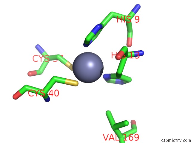
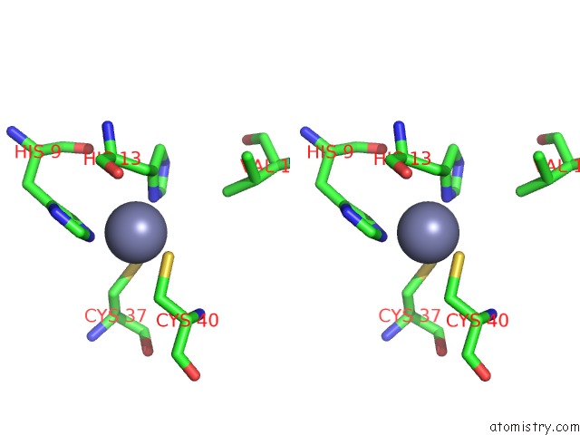
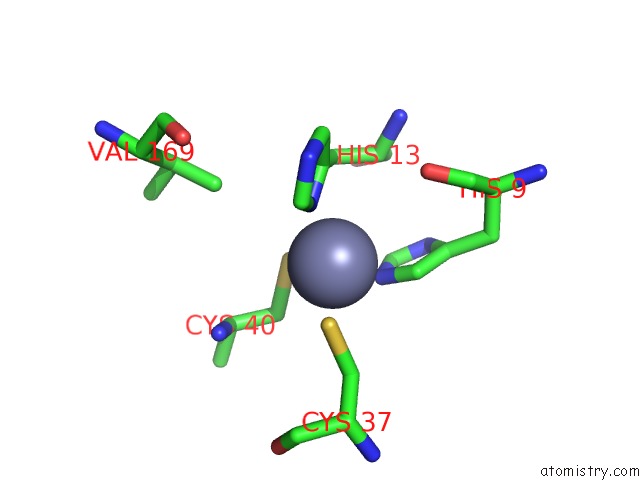
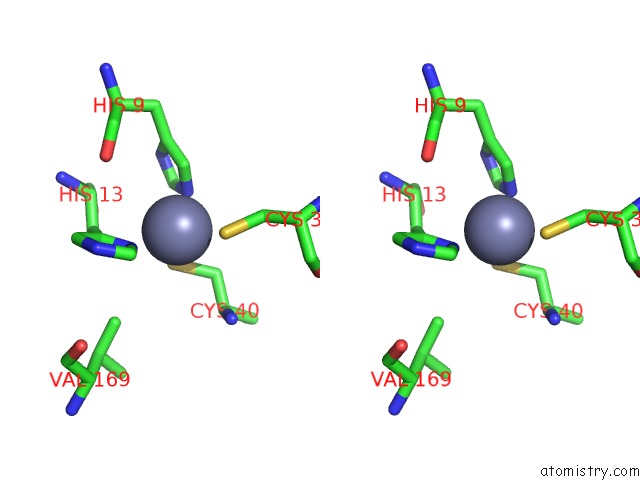
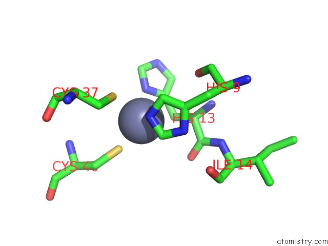
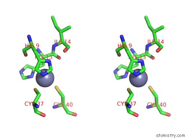
Zinc binding site 1 out of 8 in 5ejk
Go back to
Zinc binding site 1 out
of 8 in the Crystal Structure of the Rous Sarcoma Virus Intasome
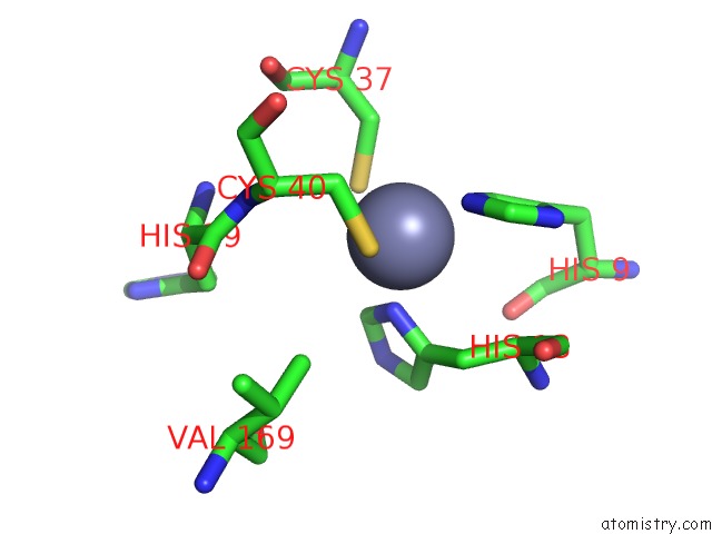
Mono view
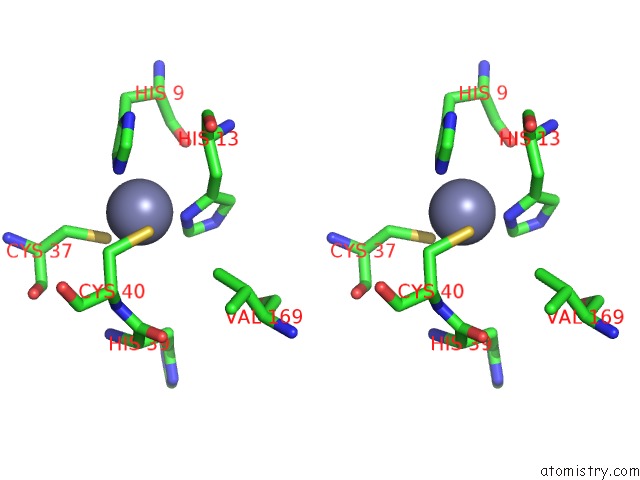
Stereo pair view
Mono view
Stereo pair view
A full contact list of Zinc with other atoms in the Zn binding
site number 1 of Crystal Structure of the Rous Sarcoma Virus Intasome within 5.0Å range:
|
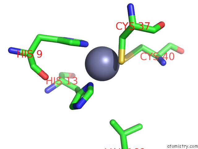
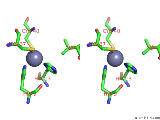
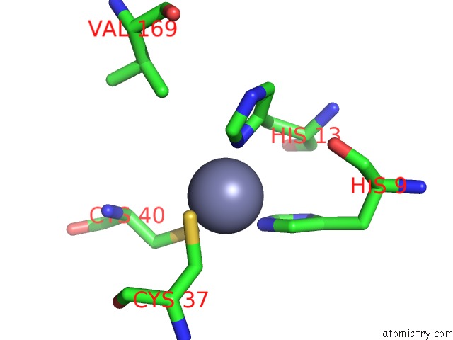
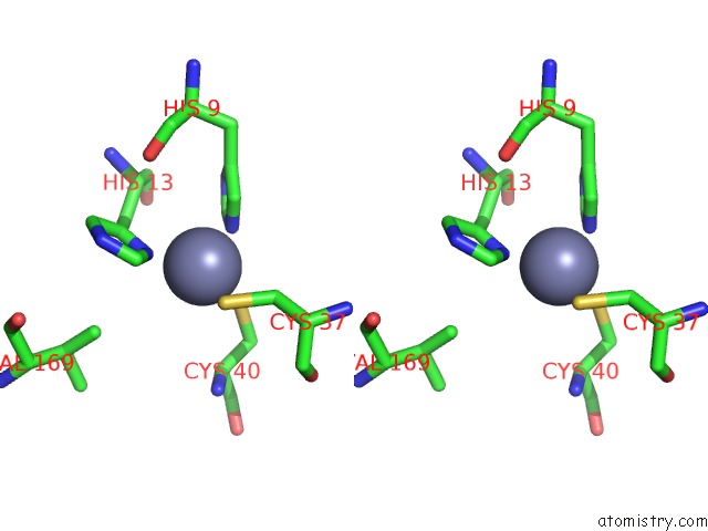
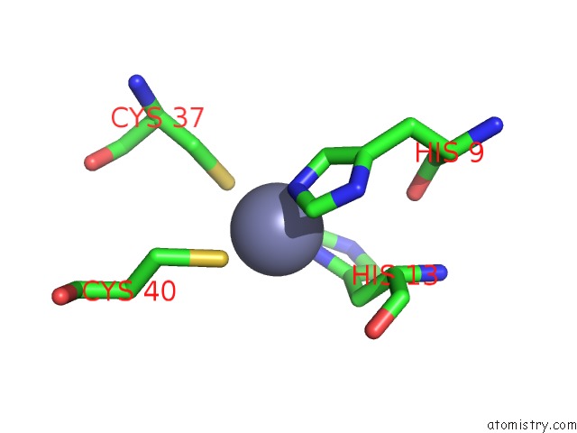
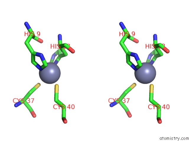
Zinc binding site 2 out of 8 in 5ejk
Go back to
Zinc binding site 2 out
of 8 in the Crystal Structure of the Rous Sarcoma Virus Intasome
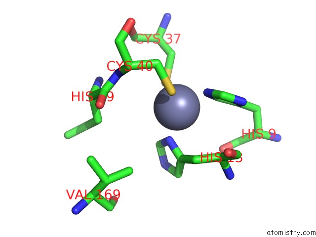
Mono view
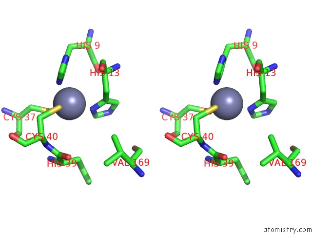
Stereo pair view
Mono view
Stereo pair view
A full contact list of Zinc with other atoms in the Zn binding
site number 2 of Crystal Structure of the Rous Sarcoma Virus Intasome within 5.0Å range:
|
Zinc binding site 3 out of 8 in 5ejk
Go back to
Zinc binding site 3 out
of 8 in the Crystal Structure of the Rous Sarcoma Virus Intasome
Mono view
Stereo pair view
Mono view
Stereo pair view
A full contact list of Zinc with other atoms in the Zn binding
site number 3 of Crystal Structure of the Rous Sarcoma Virus Intasome within 5.0Å range:
|
Zinc binding site 4 out of 8 in 5ejk
Go back to
Zinc binding site 4 out
of 8 in the Crystal Structure of the Rous Sarcoma Virus Intasome
Mono view
Stereo pair view
Mono view
Stereo pair view
A full contact list of Zinc with other atoms in the Zn binding
site number 4 of Crystal Structure of the Rous Sarcoma Virus Intasome within 5.0Å range:
|
Zinc binding site 5 out of 8 in 5ejk
Go back to
Zinc binding site 5 out
of 8 in the Crystal Structure of the Rous Sarcoma Virus Intasome
Mono view
Stereo pair view
Mono view
Stereo pair view
A full contact list of Zinc with other atoms in the Zn binding
site number 5 of Crystal Structure of the Rous Sarcoma Virus Intasome within 5.0Å range:
|
Zinc binding site 6 out of 8 in 5ejk
Go back to
Zinc binding site 6 out
of 8 in the Crystal Structure of the Rous Sarcoma Virus Intasome
Mono view
Stereo pair view
Mono view
Stereo pair view
A full contact list of Zinc with other atoms in the Zn binding
site number 6 of Crystal Structure of the Rous Sarcoma Virus Intasome within 5.0Å range:
|
Zinc binding site 7 out of 8 in 5ejk
Go back to
Zinc binding site 7 out
of 8 in the Crystal Structure of the Rous Sarcoma Virus Intasome
Mono view
Stereo pair view
Mono view
Stereo pair view
A full contact list of Zinc with other atoms in the Zn binding
site number 7 of Crystal Structure of the Rous Sarcoma Virus Intasome within 5.0Å range:
|
Zinc binding site 8 out of 8 in 5ejk
Go back to
Zinc binding site 8 out
of 8 in the Crystal Structure of the Rous Sarcoma Virus Intasome
Mono view
Stereo pair view
Mono view
Stereo pair view
A full contact list of Zinc with other atoms in the Zn binding
site number 8 of Crystal Structure of the Rous Sarcoma Virus Intasome within 5.0Å range:
|
Reference:
Z.Yin,
K.Shi,
S.Banerjee,
K.K.Pandey,
S.Bera,
D.P.Grandgenett,
H.Aihara.
Crystal Structure of the Rous Sarcoma Virus Intasome. Nature V. 530 362 2016.
ISSN: ESSN 1476-4687
PubMed: 26887497
DOI: 10.1038/NATURE16950
Page generated: Sun Oct 27 15:22:21 2024
ISSN: ESSN 1476-4687
PubMed: 26887497
DOI: 10.1038/NATURE16950
Last articles
Mo in 2VPZMo in 2VPY
Mo in 2VPX
Mo in 2VPW
Mo in 2MIN
Mo in 2V45
Mo in 2V3V
Mo in 2NYA
Mo in 2R8U
Mo in 2ONR