Zinc »
PDB 5af0-5amk »
5am9 »
Zinc in PDB 5am9: Crystal Structure of the Angiotensin-1 Converting Enzyme N- Domain in Complex with Amyloid-Beta 10-16
Enzymatic activity of Crystal Structure of the Angiotensin-1 Converting Enzyme N- Domain in Complex with Amyloid-Beta 10-16
All present enzymatic activity of Crystal Structure of the Angiotensin-1 Converting Enzyme N- Domain in Complex with Amyloid-Beta 10-16:
3.4.15.1;
3.4.15.1;
Protein crystallography data
The structure of Crystal Structure of the Angiotensin-1 Converting Enzyme N- Domain in Complex with Amyloid-Beta 10-16, PDB code: 5am9
was solved by
G.Masuyer,
K.M.Larmuth,
R.G.Douglas,
E.D.Sturrock,
K.R.Acharya,
with X-Ray Crystallography technique. A brief refinement statistics is given in the table below:
| Resolution Low / High (Å) | 113.27 / 1.80 |
| Space group | P 1 |
| Cell size a, b, c (Å), α, β, γ (°) | 73.348, 101.800, 113.950, 85.04, 85.55, 81.88 |
| R / Rfree (%) | 19.674 / 22.907 |
Other elements in 5am9:
The structure of Crystal Structure of the Angiotensin-1 Converting Enzyme N- Domain in Complex with Amyloid-Beta 10-16 also contains other interesting chemical elements:
| Calcium | (Ca) | 2 atoms |
| Chlorine | (Cl) | 4 atoms |
| Sodium | (Na) | 2 atoms |
Zinc Binding Sites:
The binding sites of Zinc atom in the Crystal Structure of the Angiotensin-1 Converting Enzyme N- Domain in Complex with Amyloid-Beta 10-16
(pdb code 5am9). This binding sites where shown within
5.0 Angstroms radius around Zinc atom.
In total 4 binding sites of Zinc where determined in the Crystal Structure of the Angiotensin-1 Converting Enzyme N- Domain in Complex with Amyloid-Beta 10-16, PDB code: 5am9:
Jump to Zinc binding site number: 1; 2; 3; 4;
In total 4 binding sites of Zinc where determined in the Crystal Structure of the Angiotensin-1 Converting Enzyme N- Domain in Complex with Amyloid-Beta 10-16, PDB code: 5am9:
Jump to Zinc binding site number: 1; 2; 3; 4;
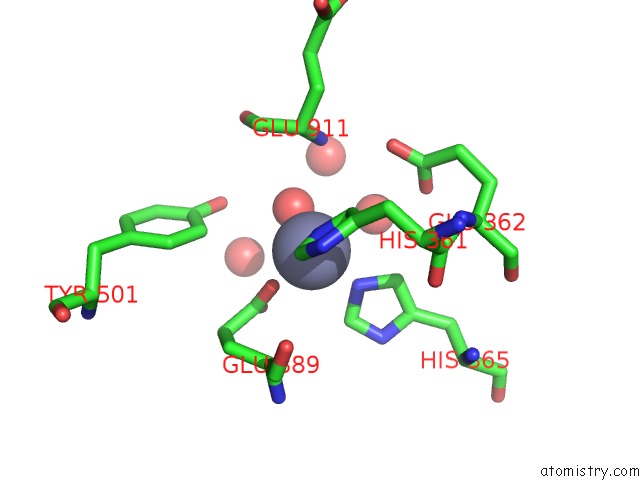
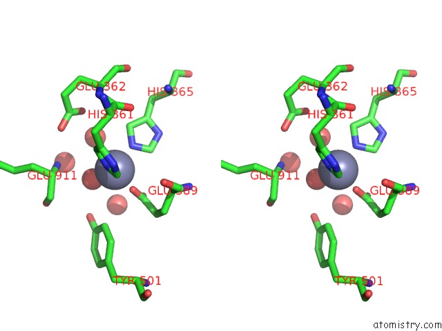
Zinc binding site 1 out of 4 in 5am9
Go back to
Zinc binding site 1 out
of 4 in the Crystal Structure of the Angiotensin-1 Converting Enzyme N- Domain in Complex with Amyloid-Beta 10-16
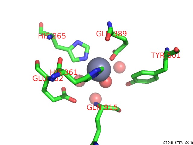
Mono view
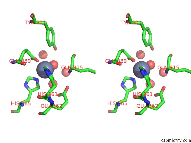
Stereo pair view
Mono view
Stereo pair view
A full contact list of Zinc with other atoms in the Zn binding
site number 1 of Crystal Structure of the Angiotensin-1 Converting Enzyme N- Domain in Complex with Amyloid-Beta 10-16 within 5.0Å range:
|
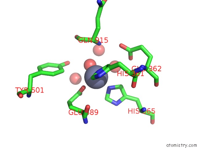
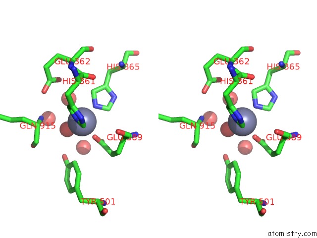
Zinc binding site 2 out of 4 in 5am9
Go back to
Zinc binding site 2 out
of 4 in the Crystal Structure of the Angiotensin-1 Converting Enzyme N- Domain in Complex with Amyloid-Beta 10-16
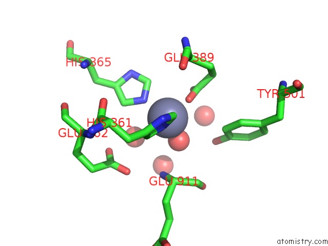
Mono view
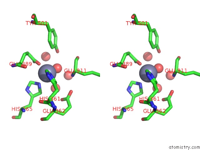
Stereo pair view
Mono view
Stereo pair view
A full contact list of Zinc with other atoms in the Zn binding
site number 2 of Crystal Structure of the Angiotensin-1 Converting Enzyme N- Domain in Complex with Amyloid-Beta 10-16 within 5.0Å range:
|
Zinc binding site 3 out of 4 in 5am9
Go back to
Zinc binding site 3 out
of 4 in the Crystal Structure of the Angiotensin-1 Converting Enzyme N- Domain in Complex with Amyloid-Beta 10-16
Mono view
Stereo pair view
Mono view
Stereo pair view
A full contact list of Zinc with other atoms in the Zn binding
site number 3 of Crystal Structure of the Angiotensin-1 Converting Enzyme N- Domain in Complex with Amyloid-Beta 10-16 within 5.0Å range:
|
Zinc binding site 4 out of 4 in 5am9
Go back to
Zinc binding site 4 out
of 4 in the Crystal Structure of the Angiotensin-1 Converting Enzyme N- Domain in Complex with Amyloid-Beta 10-16
Mono view
Stereo pair view
Mono view
Stereo pair view
A full contact list of Zinc with other atoms in the Zn binding
site number 4 of Crystal Structure of the Angiotensin-1 Converting Enzyme N- Domain in Complex with Amyloid-Beta 10-16 within 5.0Å range:
|
Reference:
K.M.Larmuth,
G.Masuyer,
R.G.Douglas,
E.D.Sturrock,
K.R.Acharya.
The Kinetic and Structural Characterisation of Amyloid-Beta Metabolism By Human Angiotensin-1- Converting Enzyme (Ace) Febs J. V. 283 1060 2016.
ISSN: ISSN 1742-464X
PubMed: 26748546
DOI: 10.1111/FEBS.13647
Page generated: Sun Oct 27 13:04:59 2024
ISSN: ISSN 1742-464X
PubMed: 26748546
DOI: 10.1111/FEBS.13647
Last articles
Mg in 4W5OMg in 4W5J
Mg in 4W5N
Mg in 4V2I
Mg in 4V3R
Mg in 4V26
Mg in 4V2G
Mg in 4V1T
Mg in 4V25
Mg in 4V1V