Zinc »
PDB 4xhv-4xo4 »
4xmz »
Zinc in PDB 4xmz: Crystal Structure of MET260ALA Mutant of E. Coli Aminopeptidase N in Complex with 2,4-Diaminobutyric Acid
Enzymatic activity of Crystal Structure of MET260ALA Mutant of E. Coli Aminopeptidase N in Complex with 2,4-Diaminobutyric Acid
All present enzymatic activity of Crystal Structure of MET260ALA Mutant of E. Coli Aminopeptidase N in Complex with 2,4-Diaminobutyric Acid:
3.4.11.2;
3.4.11.2;
Protein crystallography data
The structure of Crystal Structure of MET260ALA Mutant of E. Coli Aminopeptidase N in Complex with 2,4-Diaminobutyric Acid, PDB code: 4xmz
was solved by
A.Addlagatta,
R.Gumpena,
with X-Ray Crystallography technique. A brief refinement statistics is given in the table below:
| Resolution Low / High (Å) | 19.81 / 2.15 |
| Space group | P 31 2 1 |
| Cell size a, b, c (Å), α, β, γ (°) | 120.638, 120.638, 171.304, 90.00, 90.00, 120.00 |
| R / Rfree (%) | 14.3 / 18.1 |
Other elements in 4xmz:
The structure of Crystal Structure of MET260ALA Mutant of E. Coli Aminopeptidase N in Complex with 2,4-Diaminobutyric Acid also contains other interesting chemical elements:
| Sodium | (Na) | 8 atoms |
Zinc Binding Sites:
The binding sites of Zinc atom in the Crystal Structure of MET260ALA Mutant of E. Coli Aminopeptidase N in Complex with 2,4-Diaminobutyric Acid
(pdb code 4xmz). This binding sites where shown within
5.0 Angstroms radius around Zinc atom.
In total only one binding site of Zinc was determined in the Crystal Structure of MET260ALA Mutant of E. Coli Aminopeptidase N in Complex with 2,4-Diaminobutyric Acid, PDB code: 4xmz:
In total only one binding site of Zinc was determined in the Crystal Structure of MET260ALA Mutant of E. Coli Aminopeptidase N in Complex with 2,4-Diaminobutyric Acid, PDB code: 4xmz:
Zinc binding site 1 out of 1 in 4xmz
Go back to
Zinc binding site 1 out
of 1 in the Crystal Structure of MET260ALA Mutant of E. Coli Aminopeptidase N in Complex with 2,4-Diaminobutyric Acid
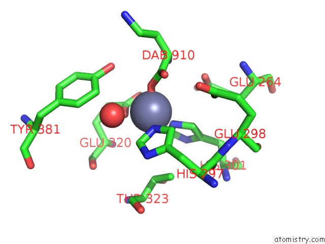
Mono view
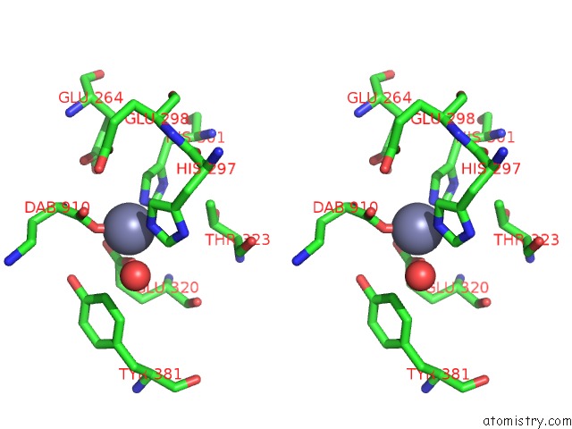
Stereo pair view
Mono view
Stereo pair view
A full contact list of Zinc with other atoms in the Zn binding
site number 1 of Crystal Structure of MET260ALA Mutant of E. Coli Aminopeptidase N in Complex with 2,4-Diaminobutyric Acid within 5.0Å range:
|
Reference:
A.Addlagatta,
R.Gumpena.
Crystal Structure of MET260ALA Mutant of E. Coli Aminopeptidase N in Complex with 2,4-Diaminobutyric Acid To Be Published.
Page generated: Sun Oct 27 10:41:17 2024
Last articles
Mn in 5NRZMn in 5NMP
Mn in 5NHA
Mn in 5NPK
Mn in 5NPP
Mn in 5NNB
Mn in 5NNA
Mn in 5NH9
Mn in 5NDF
Mn in 5NFN