Zinc »
PDB 4wnv-4x3r »
4wpv »
Zinc in PDB 4wpv: Crystal Structure of E83A Mutant of Mtb Pepck in Complex with ZN2+ and Phosphate Ion
Enzymatic activity of Crystal Structure of E83A Mutant of Mtb Pepck in Complex with ZN2+ and Phosphate Ion
All present enzymatic activity of Crystal Structure of E83A Mutant of Mtb Pepck in Complex with ZN2+ and Phosphate Ion:
4.1.1.32;
4.1.1.32;
Protein crystallography data
The structure of Crystal Structure of E83A Mutant of Mtb Pepck in Complex with ZN2+ and Phosphate Ion, PDB code: 4wpv
was solved by
H.L.Kim,
J.C.Sacchettini,
with X-Ray Crystallography technique. A brief refinement statistics is given in the table below:
| Resolution Low / High (Å) | 47.81 / 1.67 |
| Space group | C 1 2 1 |
| Cell size a, b, c (Å), α, β, γ (°) | 102.762, 122.281, 63.984, 90.00, 116.88, 90.00 |
| R / Rfree (%) | 15.5 / 17.7 |
Zinc Binding Sites:
The binding sites of Zinc atom in the Crystal Structure of E83A Mutant of Mtb Pepck in Complex with ZN2+ and Phosphate Ion
(pdb code 4wpv). This binding sites where shown within
5.0 Angstroms radius around Zinc atom.
In total 2 binding sites of Zinc where determined in the Crystal Structure of E83A Mutant of Mtb Pepck in Complex with ZN2+ and Phosphate Ion, PDB code: 4wpv:
Jump to Zinc binding site number: 1; 2;
In total 2 binding sites of Zinc where determined in the Crystal Structure of E83A Mutant of Mtb Pepck in Complex with ZN2+ and Phosphate Ion, PDB code: 4wpv:
Jump to Zinc binding site number: 1; 2;
Zinc binding site 1 out of 2 in 4wpv
Go back to
Zinc binding site 1 out
of 2 in the Crystal Structure of E83A Mutant of Mtb Pepck in Complex with ZN2+ and Phosphate Ion
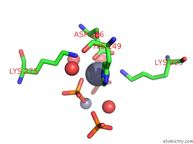
Mono view
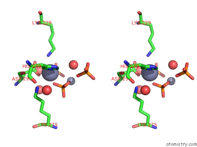
Stereo pair view
Mono view
Stereo pair view
A full contact list of Zinc with other atoms in the Zn binding
site number 1 of Crystal Structure of E83A Mutant of Mtb Pepck in Complex with ZN2+ and Phosphate Ion within 5.0Å range:
|
Zinc binding site 2 out of 2 in 4wpv
Go back to
Zinc binding site 2 out
of 2 in the Crystal Structure of E83A Mutant of Mtb Pepck in Complex with ZN2+ and Phosphate Ion
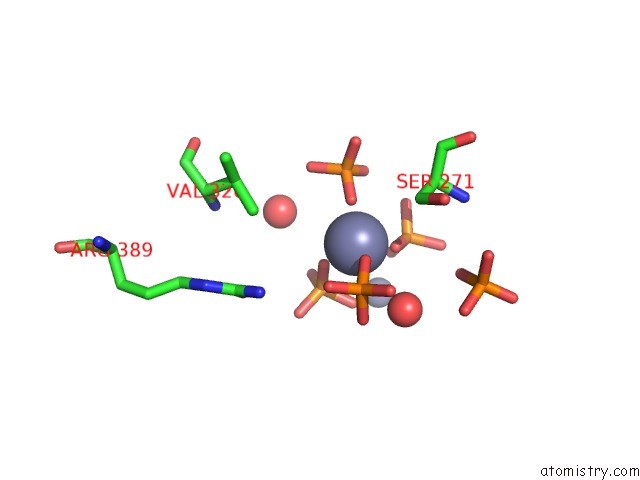
Mono view
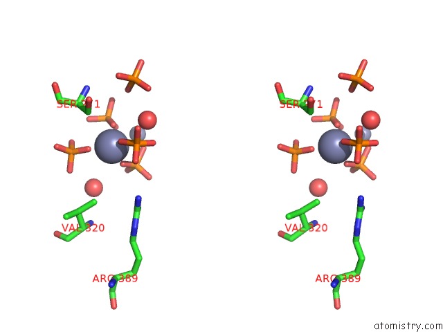
Stereo pair view
Mono view
Stereo pair view
A full contact list of Zinc with other atoms in the Zn binding
site number 2 of Crystal Structure of E83A Mutant of Mtb Pepck in Complex with ZN2+ and Phosphate Ion within 5.0Å range:
|
Reference:
H.L.Kim,
J.C.Sacchettini.
Crystal Structure of E83A From Mtb Pepck in Complex with ZN2+ and Phosphate Ion To Be Published.
Page generated: Sun Oct 27 10:01:57 2024
Last articles
I in 3SEXI in 3SLS
I in 3SKQ
I in 3SKF
I in 3SJF
I in 3S1S
I in 3S99
I in 3SD0
I in 3RU6
I in 3S43