Zinc »
PDB 4tpm-4u6a »
4tsd »
Zinc in PDB 4tsd: Crystal Structure of Helicobacter Pylori HP1029
Protein crystallography data
The structure of Crystal Structure of Helicobacter Pylori HP1029, PDB code: 4tsd
was solved by
F.Vallese,
R.Percudani,
G.Zanotti,
with X-Ray Crystallography technique. A brief refinement statistics is given in the table below:
| Resolution Low / High (Å) | 49.57 / 1.53 |
| Space group | P 21 21 21 |
| Cell size a, b, c (Å), α, β, γ (°) | 44.788, 77.490, 99.134, 90.00, 90.00, 90.00 |
| R / Rfree (%) | 15.8 / 18.5 |
Zinc Binding Sites:
The binding sites of Zinc atom in the Crystal Structure of Helicobacter Pylori HP1029
(pdb code 4tsd). This binding sites where shown within
5.0 Angstroms radius around Zinc atom.
In total 2 binding sites of Zinc where determined in the Crystal Structure of Helicobacter Pylori HP1029, PDB code: 4tsd:
Jump to Zinc binding site number: 1; 2;
In total 2 binding sites of Zinc where determined in the Crystal Structure of Helicobacter Pylori HP1029, PDB code: 4tsd:
Jump to Zinc binding site number: 1; 2;
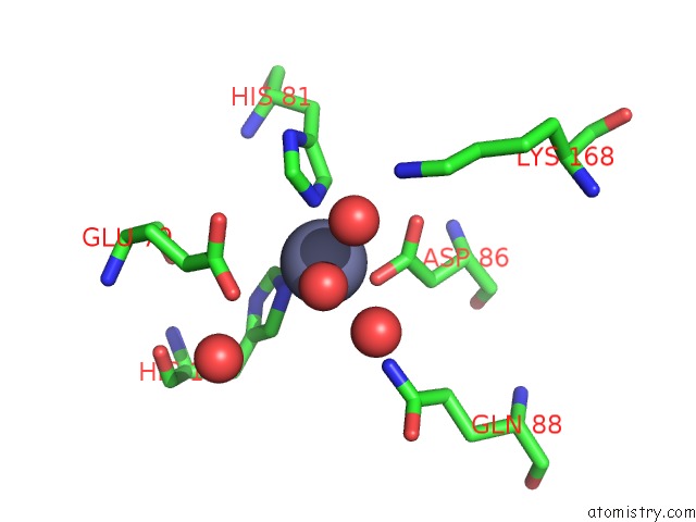
Zinc binding site 1 out of 2 in 4tsd
Go back to
Zinc binding site 1 out
of 2 in the Crystal Structure of Helicobacter Pylori HP1029

Mono view
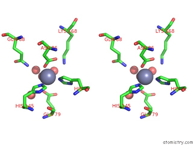
Stereo pair view
Mono view
Stereo pair view
A full contact list of Zinc with other atoms in the Zn binding
site number 1 of Crystal Structure of Helicobacter Pylori HP1029 within 5.0Å range:
|
Zinc binding site 2 out of 2 in 4tsd
Go back to
Zinc binding site 2 out
of 2 in the Crystal Structure of Helicobacter Pylori HP1029
Mono view
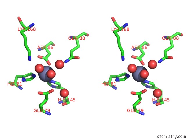
Stereo pair view
Mono view
Stereo pair view
A full contact list of Zinc with other atoms in the Zn binding
site number 2 of Crystal Structure of Helicobacter Pylori HP1029 within 5.0Å range:
|
Reference:
F.Vallese,
R.Percudani,
W.Fischer,
G.Zanotti.
The Crystal Structure of Helicobacter Pylori HP1029 Highlights the Functional Diversity of the Sialic Acid-Related DUF386 Family. Febs J. V. 282 3311 2015.
ISSN: ISSN 1742-464X
PubMed: 26096900
DOI: 10.1111/FEBS.13344
Page generated: Sun Oct 27 08:34:46 2024
ISSN: ISSN 1742-464X
PubMed: 26096900
DOI: 10.1111/FEBS.13344
Last articles
Mg in 9GUPMg in 9GUR
Mg in 9GRE
Mg in 9GTK
Mg in 9GU5
Mg in 9GOB
Mg in 9GO5
Mg in 9GMZ
Mg in 9GQO
Mg in 9GMX