Zinc »
PDB 4rvo-4tpj »
4s1z »
Zinc in PDB 4s1z: Crystal Structure of Trabid NZF1 in Complex with K29 Linked Di- Ubiquitin
Enzymatic activity of Crystal Structure of Trabid NZF1 in Complex with K29 Linked Di- Ubiquitin
All present enzymatic activity of Crystal Structure of Trabid NZF1 in Complex with K29 Linked Di- Ubiquitin:
3.4.19.12;
3.4.19.12;
Protein crystallography data
The structure of Crystal Structure of Trabid NZF1 in Complex with K29 Linked Di- Ubiquitin, PDB code: 4s1z
was solved by
Y.A.Kristariyanto,
S.A.Abdul Rehman,
D.G.Campbell,
N.A.Morrice,
C.Johnson,
R.Toth,
Y.Kulathu,
with X-Ray Crystallography technique. A brief refinement statistics is given in the table below:
| Resolution Low / High (Å) | 76.10 / 3.03 |
| Space group | C 1 2 1 |
| Cell size a, b, c (Å), α, β, γ (°) | 99.222, 123.971, 78.312, 90.00, 103.68, 90.00 |
| R / Rfree (%) | 22.2 / 27 |
Zinc Binding Sites:
The binding sites of Zinc atom in the Crystal Structure of Trabid NZF1 in Complex with K29 Linked Di- Ubiquitin
(pdb code 4s1z). This binding sites where shown within
5.0 Angstroms radius around Zinc atom.
In total 5 binding sites of Zinc where determined in the Crystal Structure of Trabid NZF1 in Complex with K29 Linked Di- Ubiquitin, PDB code: 4s1z:
Jump to Zinc binding site number: 1; 2; 3; 4; 5;
In total 5 binding sites of Zinc where determined in the Crystal Structure of Trabid NZF1 in Complex with K29 Linked Di- Ubiquitin, PDB code: 4s1z:
Jump to Zinc binding site number: 1; 2; 3; 4; 5;
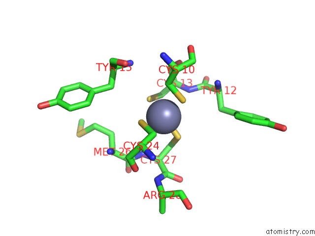
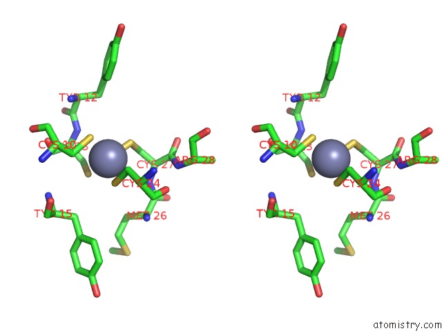
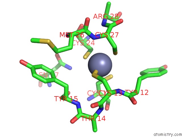
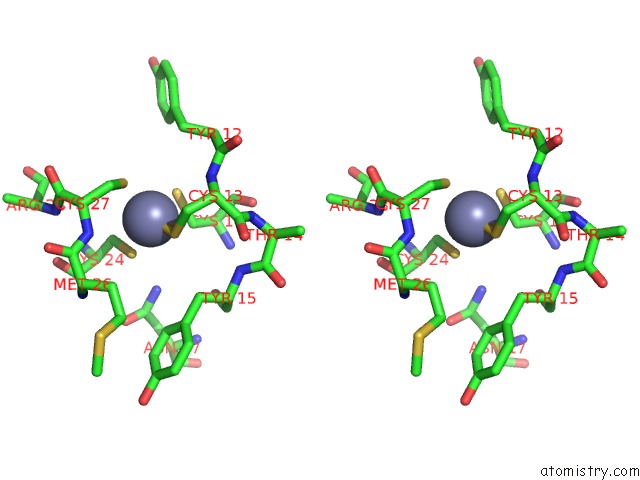
Zinc binding site 1 out of 5 in 4s1z
Go back to
Zinc binding site 1 out
of 5 in the Crystal Structure of Trabid NZF1 in Complex with K29 Linked Di- Ubiquitin
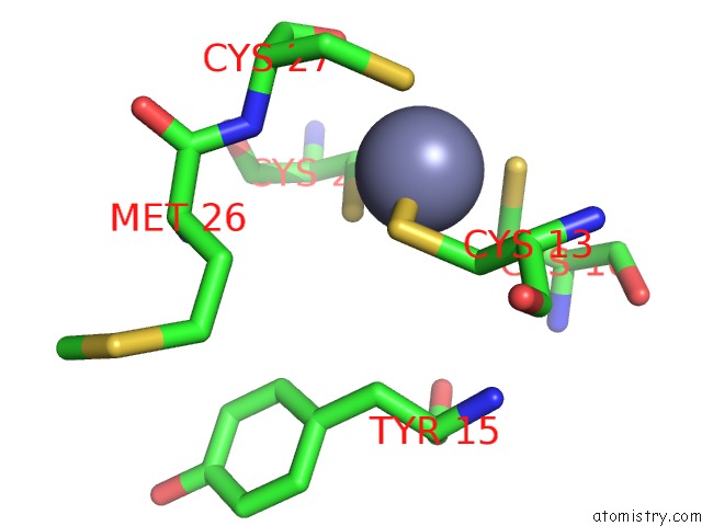
Mono view
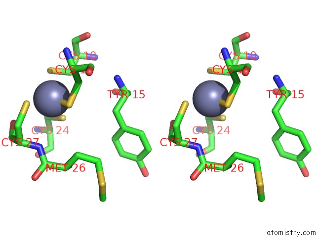
Stereo pair view
Mono view
Stereo pair view
A full contact list of Zinc with other atoms in the Zn binding
site number 1 of Crystal Structure of Trabid NZF1 in Complex with K29 Linked Di- Ubiquitin within 5.0Å range:
|
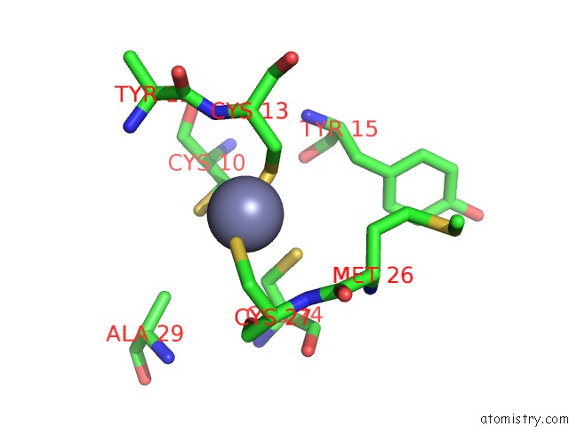
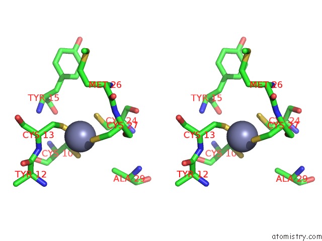
Zinc binding site 2 out of 5 in 4s1z
Go back to
Zinc binding site 2 out
of 5 in the Crystal Structure of Trabid NZF1 in Complex with K29 Linked Di- Ubiquitin
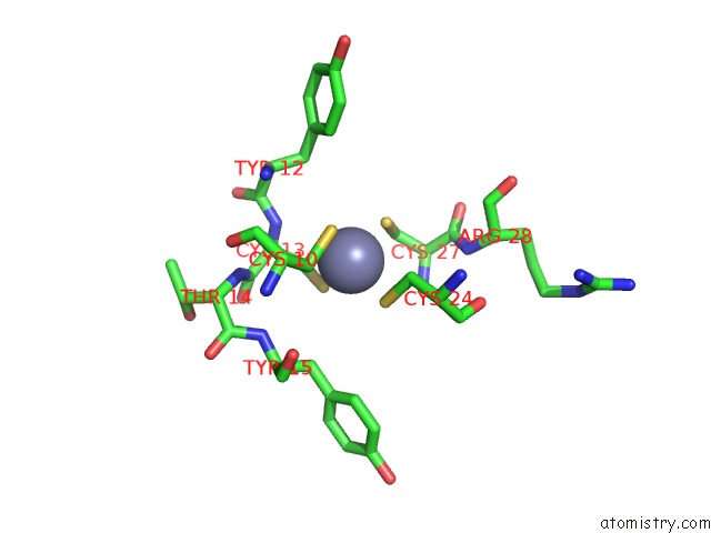
Mono view
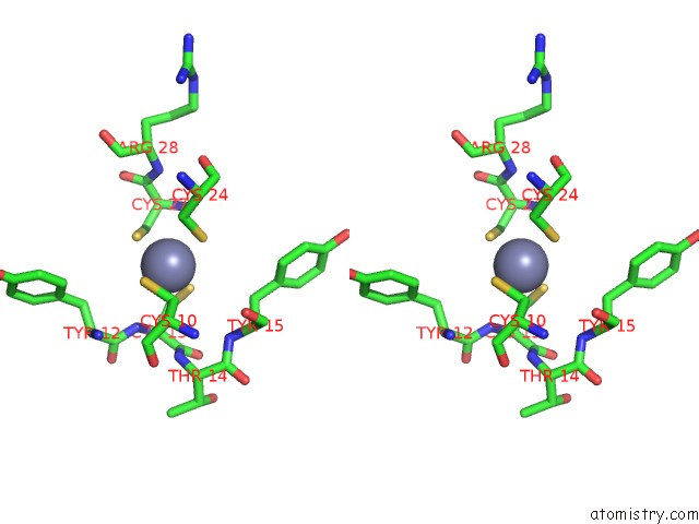
Stereo pair view
Mono view
Stereo pair view
A full contact list of Zinc with other atoms in the Zn binding
site number 2 of Crystal Structure of Trabid NZF1 in Complex with K29 Linked Di- Ubiquitin within 5.0Å range:
|
Zinc binding site 3 out of 5 in 4s1z
Go back to
Zinc binding site 3 out
of 5 in the Crystal Structure of Trabid NZF1 in Complex with K29 Linked Di- Ubiquitin
Mono view
Stereo pair view
Mono view
Stereo pair view
A full contact list of Zinc with other atoms in the Zn binding
site number 3 of Crystal Structure of Trabid NZF1 in Complex with K29 Linked Di- Ubiquitin within 5.0Å range:
|
Zinc binding site 4 out of 5 in 4s1z
Go back to
Zinc binding site 4 out
of 5 in the Crystal Structure of Trabid NZF1 in Complex with K29 Linked Di- Ubiquitin
Mono view
Stereo pair view
Mono view
Stereo pair view
A full contact list of Zinc with other atoms in the Zn binding
site number 4 of Crystal Structure of Trabid NZF1 in Complex with K29 Linked Di- Ubiquitin within 5.0Å range:
|
Zinc binding site 5 out of 5 in 4s1z
Go back to
Zinc binding site 5 out
of 5 in the Crystal Structure of Trabid NZF1 in Complex with K29 Linked Di- Ubiquitin
Mono view
Stereo pair view
Mono view
Stereo pair view
A full contact list of Zinc with other atoms in the Zn binding
site number 5 of Crystal Structure of Trabid NZF1 in Complex with K29 Linked Di- Ubiquitin within 5.0Å range:
|
Reference:
Y.A.Kristariyanto,
S.A.Abdul Rehman,
D.G.Campbell,
N.A.Morrice,
C.Johnson,
R.Toth,
Y.Kulathu.
K29-Selective Ubiquitin Binding Domain Reveals Structural Basis of Specificity and Heterotypic Nature of K29 Polyubiquitin. Mol.Cell 2015.
ISSN: ISSN 1097-2765
PubMed: 25752573
DOI: 10.1016/J.MOLCEL.2015.01.041
Page generated: Sun Oct 27 07:27:11 2024
ISSN: ISSN 1097-2765
PubMed: 25752573
DOI: 10.1016/J.MOLCEL.2015.01.041
Last articles
Mg in 9GURMg in 9GRE
Mg in 9GTK
Mg in 9GU5
Mg in 9GOB
Mg in 9GO5
Mg in 9GMZ
Mg in 9GQO
Mg in 9GMX
Mg in 9GMW