Zinc »
PDB 4o8x-4ojv »
4ocm »
Zinc in PDB 4ocm: Crystal Structure of the RPN8-RPN11 Mpn Domain Heterodimer, Crystal Form Ib
Protein crystallography data
The structure of Crystal Structure of the RPN8-RPN11 Mpn Domain Heterodimer, Crystal Form Ib, PDB code: 4ocm
was solved by
G.R.Pathare,
A.Bracher,
with X-Ray Crystallography technique. A brief refinement statistics is given in the table below:
| Resolution Low / High (Å) | 30.00 / 1.99 |
| Space group | P 1 2 1 |
| Cell size a, b, c (Å), α, β, γ (°) | 63.401, 44.970, 200.043, 90.00, 98.40, 90.00 |
| R / Rfree (%) | 21.6 / 26.2 |
Other elements in 4ocm:
The structure of Crystal Structure of the RPN8-RPN11 Mpn Domain Heterodimer, Crystal Form Ib also contains other interesting chemical elements:
| Potassium | (K) | 1 atom |
Zinc Binding Sites:
The binding sites of Zinc atom in the Crystal Structure of the RPN8-RPN11 Mpn Domain Heterodimer, Crystal Form Ib
(pdb code 4ocm). This binding sites where shown within
5.0 Angstroms radius around Zinc atom.
In total 2 binding sites of Zinc where determined in the Crystal Structure of the RPN8-RPN11 Mpn Domain Heterodimer, Crystal Form Ib, PDB code: 4ocm:
Jump to Zinc binding site number: 1; 2;
In total 2 binding sites of Zinc where determined in the Crystal Structure of the RPN8-RPN11 Mpn Domain Heterodimer, Crystal Form Ib, PDB code: 4ocm:
Jump to Zinc binding site number: 1; 2;
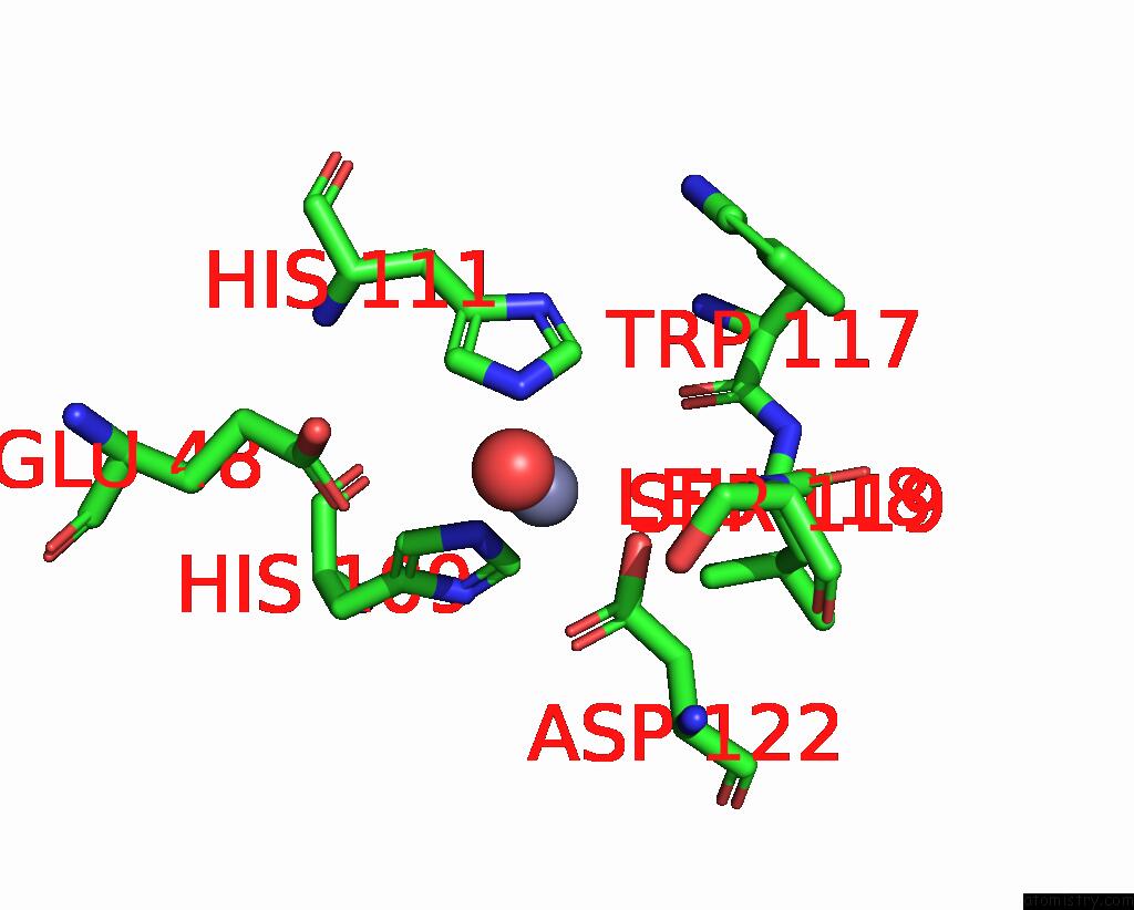
Zinc binding site 1 out of 2 in 4ocm
Go back to
Zinc binding site 1 out
of 2 in the Crystal Structure of the RPN8-RPN11 Mpn Domain Heterodimer, Crystal Form Ib

Mono view
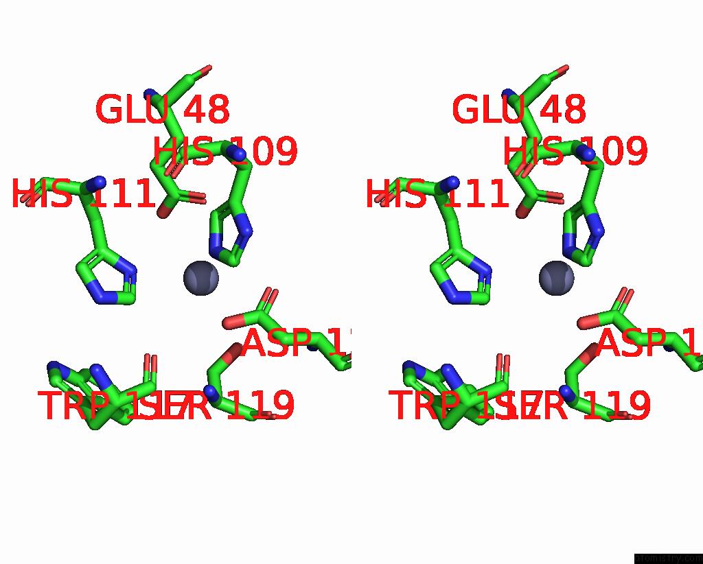
Stereo pair view
Mono view
Stereo pair view
A full contact list of Zinc with other atoms in the Zn binding
site number 1 of Crystal Structure of the RPN8-RPN11 Mpn Domain Heterodimer, Crystal Form Ib within 5.0Å range:
|
Zinc binding site 2 out of 2 in 4ocm
Go back to
Zinc binding site 2 out
of 2 in the Crystal Structure of the RPN8-RPN11 Mpn Domain Heterodimer, Crystal Form Ib
Mono view
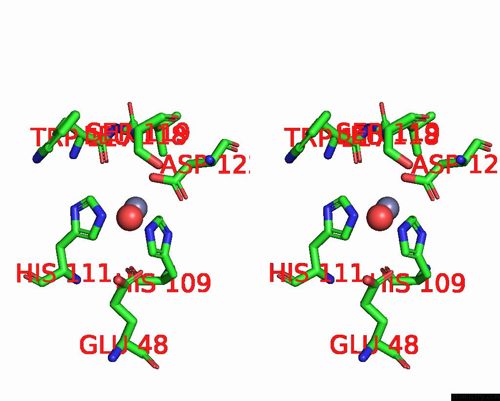
Stereo pair view
Mono view
Stereo pair view
A full contact list of Zinc with other atoms in the Zn binding
site number 2 of Crystal Structure of the RPN8-RPN11 Mpn Domain Heterodimer, Crystal Form Ib within 5.0Å range:
|
Reference:
G.R.Pathare,
I.Nagy,
P.Sledz,
D.J.Anderson,
H.J.Zhou,
E.Pardon,
J.Steyaert,
F.Forster,
A.Bracher,
W.Baumeister.
Crystal Structure of the Proteasomal Deubiquitylation Module RPN8-RPN11. Proc.Natl.Acad.Sci.Usa V. 111 2984 2014.
ISSN: ISSN 0027-8424
PubMed: 24516147
DOI: 10.1073/PNAS.1400546111
Page generated: Sun Oct 27 03:40:01 2024
ISSN: ISSN 0027-8424
PubMed: 24516147
DOI: 10.1073/PNAS.1400546111
Last articles
I in 3SKQI in 3SKF
I in 3SJF
I in 3S1S
I in 3S99
I in 3SD0
I in 3RU6
I in 3S43
I in 3S5Q
I in 3S53