Zinc »
PDB 4m65-4mi5 »
4mhn »
Zinc in PDB 4mhn: Crystal Structure of A Glutaminyl Cyclase From Ixodes Scapularis
Enzymatic activity of Crystal Structure of A Glutaminyl Cyclase From Ixodes Scapularis
All present enzymatic activity of Crystal Structure of A Glutaminyl Cyclase From Ixodes Scapularis:
2.3.2.5;
2.3.2.5;
Protein crystallography data
The structure of Crystal Structure of A Glutaminyl Cyclase From Ixodes Scapularis, PDB code: 4mhn
was solved by
K.F.Huang,
H.L.Hsu,
A.H.J.Wang,
with X-Ray Crystallography technique. A brief refinement statistics is given in the table below:
| Resolution Low / High (Å) | 30.00 / 1.15 |
| Space group | P 21 21 21 |
| Cell size a, b, c (Å), α, β, γ (°) | 55.278, 71.379, 80.076, 90.00, 90.00, 90.00 |
| R / Rfree (%) | 16.2 / 18.1 |
Zinc Binding Sites:
The binding sites of Zinc atom in the Crystal Structure of A Glutaminyl Cyclase From Ixodes Scapularis
(pdb code 4mhn). This binding sites where shown within
5.0 Angstroms radius around Zinc atom.
In total only one binding site of Zinc was determined in the Crystal Structure of A Glutaminyl Cyclase From Ixodes Scapularis, PDB code: 4mhn:
In total only one binding site of Zinc was determined in the Crystal Structure of A Glutaminyl Cyclase From Ixodes Scapularis, PDB code: 4mhn:
Zinc binding site 1 out of 1 in 4mhn
Go back to
Zinc binding site 1 out
of 1 in the Crystal Structure of A Glutaminyl Cyclase From Ixodes Scapularis
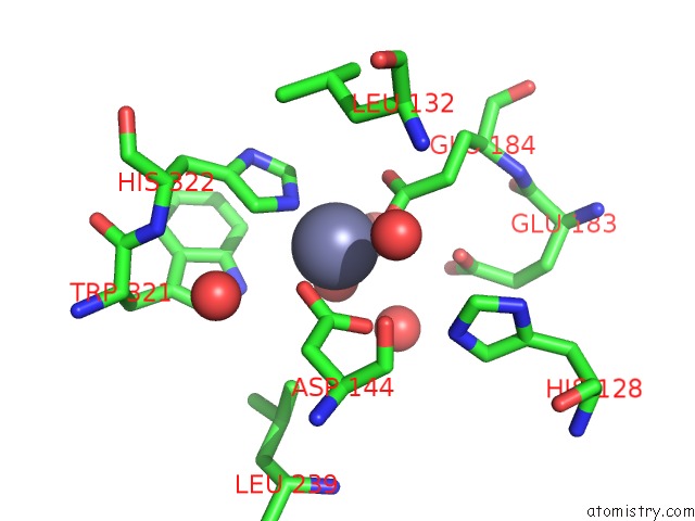
Mono view
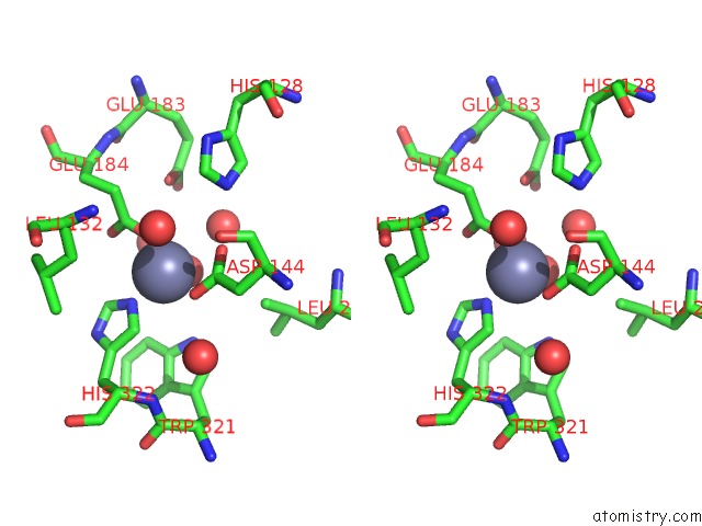
Stereo pair view
Mono view
Stereo pair view
A full contact list of Zinc with other atoms in the Zn binding
site number 1 of Crystal Structure of A Glutaminyl Cyclase From Ixodes Scapularis within 5.0Å range:
|
Reference:
K.F.Huang,
H.L.Hsu,
S.Karim,
A.H.J.Wang.
Structural and Functional Analyses of A Glutaminyl Cyclase From Ixodes Scapularis Reveal Metal-Independent Catalysis and Inhibitor Binding. Acta Crystallogr.,Sect.D V. 70 789 2014.
ISSN: ISSN 0907-4449
DOI: 10.1107/S1399004713033488
Page generated: Wed Aug 20 20:23:16 2025
ISSN: ISSN 0907-4449
DOI: 10.1107/S1399004713033488
Last articles
K in 9NESK in 9PHG
K in 9NEI
K in 9NED
K in 9NEC
K in 9NEG
K in 9CWU
K in 9CVB
K in 9CVA
K in 9COM