Zinc »
PDB 4m3p-4mi0 »
4mec »
Zinc in PDB 4mec: Crystal Structure of Rat Heme Oxygenase-1 in Complex with Zn(II)- Protoporphyrin IX
Enzymatic activity of Crystal Structure of Rat Heme Oxygenase-1 in Complex with Zn(II)- Protoporphyrin IX
All present enzymatic activity of Crystal Structure of Rat Heme Oxygenase-1 in Complex with Zn(II)- Protoporphyrin IX:
1.14.99.3;
1.14.99.3;
Protein crystallography data
The structure of Crystal Structure of Rat Heme Oxygenase-1 in Complex with Zn(II)- Protoporphyrin IX, PDB code: 4mec
was solved by
M.Sugishima,
with X-Ray Crystallography technique. A brief refinement statistics is given in the table below:
| Resolution Low / High (Å) | 38.94 / 3.20 |
| Space group | P 1 |
| Cell size a, b, c (Å), α, β, γ (°) | 40.059, 73.167, 148.757, 86.41, 87.62, 86.25 |
| R / Rfree (%) | 24.9 / 29.9 |
Zinc Binding Sites:
The binding sites of Zinc atom in the Crystal Structure of Rat Heme Oxygenase-1 in Complex with Zn(II)- Protoporphyrin IX
(pdb code 4mec). This binding sites where shown within
5.0 Angstroms radius around Zinc atom.
In total 7 binding sites of Zinc where determined in the Crystal Structure of Rat Heme Oxygenase-1 in Complex with Zn(II)- Protoporphyrin IX, PDB code: 4mec:
Jump to Zinc binding site number: 1; 2; 3; 4; 5; 6; 7;
In total 7 binding sites of Zinc where determined in the Crystal Structure of Rat Heme Oxygenase-1 in Complex with Zn(II)- Protoporphyrin IX, PDB code: 4mec:
Jump to Zinc binding site number: 1; 2; 3; 4; 5; 6; 7;
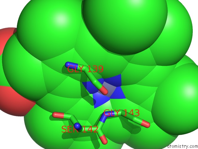
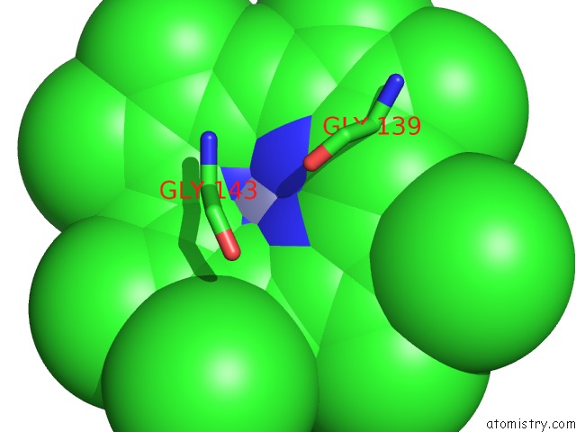
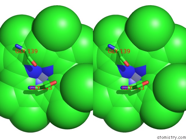
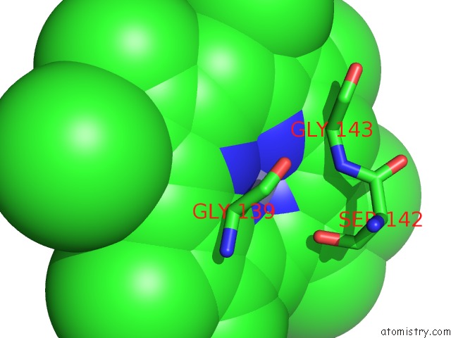
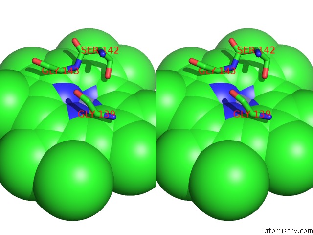
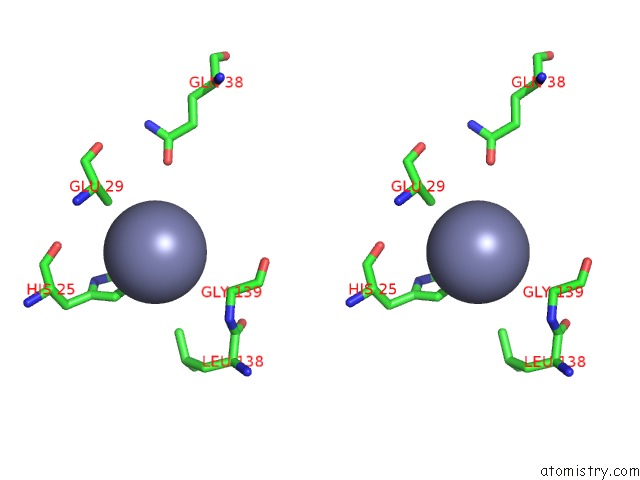
Zinc binding site 1 out of 7 in 4mec
Go back to
Zinc binding site 1 out
of 7 in the Crystal Structure of Rat Heme Oxygenase-1 in Complex with Zn(II)- Protoporphyrin IX
Mono view
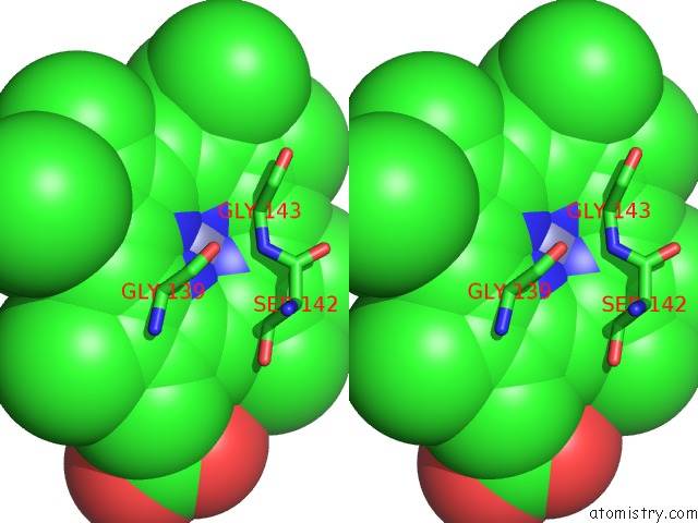
Stereo pair view
Mono view
Stereo pair view
A full contact list of Zinc with other atoms in the Zn binding
site number 1 of Crystal Structure of Rat Heme Oxygenase-1 in Complex with Zn(II)- Protoporphyrin IX within 5.0Å range:
|
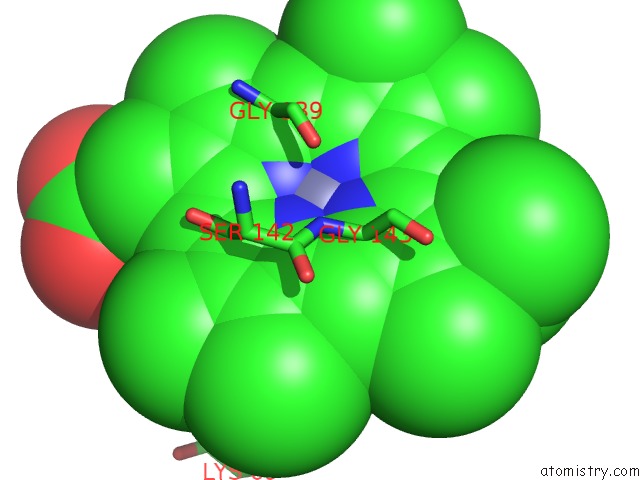
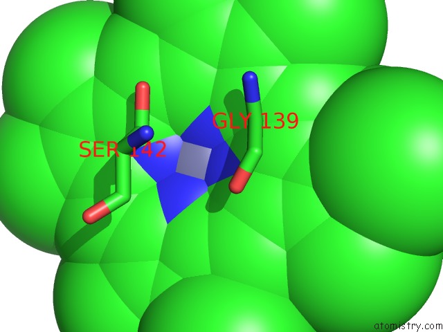
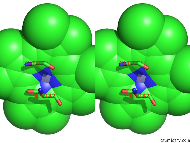
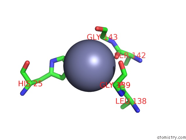
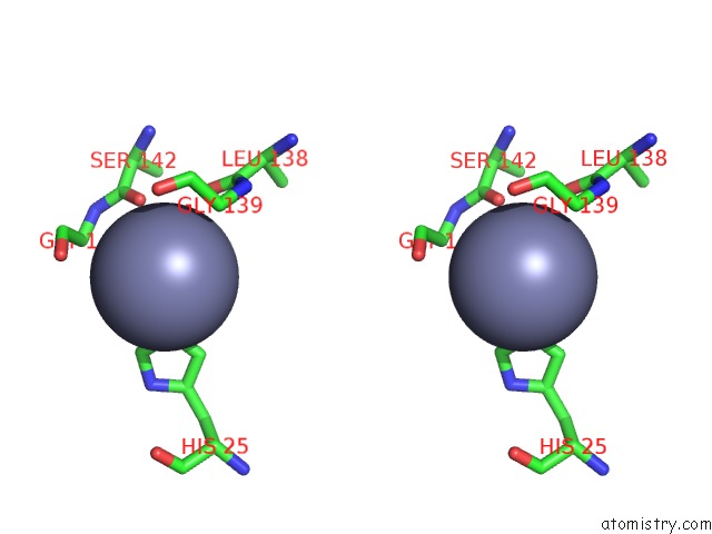
Zinc binding site 2 out of 7 in 4mec
Go back to
Zinc binding site 2 out
of 7 in the Crystal Structure of Rat Heme Oxygenase-1 in Complex with Zn(II)- Protoporphyrin IX
Mono view
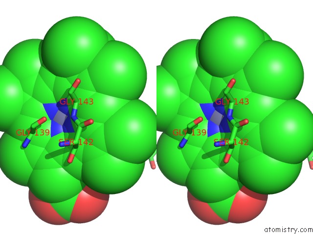
Stereo pair view
Mono view
Stereo pair view
A full contact list of Zinc with other atoms in the Zn binding
site number 2 of Crystal Structure of Rat Heme Oxygenase-1 in Complex with Zn(II)- Protoporphyrin IX within 5.0Å range:
|
Zinc binding site 3 out of 7 in 4mec
Go back to
Zinc binding site 3 out
of 7 in the Crystal Structure of Rat Heme Oxygenase-1 in Complex with Zn(II)- Protoporphyrin IX
Mono view
Stereo pair view
Mono view
Stereo pair view
A full contact list of Zinc with other atoms in the Zn binding
site number 3 of Crystal Structure of Rat Heme Oxygenase-1 in Complex with Zn(II)- Protoporphyrin IX within 5.0Å range:
|
Zinc binding site 4 out of 7 in 4mec
Go back to
Zinc binding site 4 out
of 7 in the Crystal Structure of Rat Heme Oxygenase-1 in Complex with Zn(II)- Protoporphyrin IX
Mono view
Stereo pair view
Mono view
Stereo pair view
A full contact list of Zinc with other atoms in the Zn binding
site number 4 of Crystal Structure of Rat Heme Oxygenase-1 in Complex with Zn(II)- Protoporphyrin IX within 5.0Å range:
|
Zinc binding site 5 out of 7 in 4mec
Go back to
Zinc binding site 5 out
of 7 in the Crystal Structure of Rat Heme Oxygenase-1 in Complex with Zn(II)- Protoporphyrin IX
Mono view
Stereo pair view
Mono view
Stereo pair view
A full contact list of Zinc with other atoms in the Zn binding
site number 5 of Crystal Structure of Rat Heme Oxygenase-1 in Complex with Zn(II)- Protoporphyrin IX within 5.0Å range:
|
Zinc binding site 6 out of 7 in 4mec
Go back to
Zinc binding site 6 out
of 7 in the Crystal Structure of Rat Heme Oxygenase-1 in Complex with Zn(II)- Protoporphyrin IX
Mono view
Stereo pair view
Mono view
Stereo pair view
A full contact list of Zinc with other atoms in the Zn binding
site number 6 of Crystal Structure of Rat Heme Oxygenase-1 in Complex with Zn(II)- Protoporphyrin IX within 5.0Å range:
|
Zinc binding site 7 out of 7 in 4mec
Go back to
Zinc binding site 7 out
of 7 in the Crystal Structure of Rat Heme Oxygenase-1 in Complex with Zn(II)- Protoporphyrin IX

Mono view
Stereo pair view
Mono view
Stereo pair view
A full contact list of Zinc with other atoms in the Zn binding
site number 7 of Crystal Structure of Rat Heme Oxygenase-1 in Complex with Zn(II)- Protoporphyrin IX within 5.0Å range:
|
Reference:
E.Harada,
M.Sugishima,
J.Harada,
M.Noguchi,
K.Fukuyama,
K.Sugase.
Mechanism of the Distal Regulation of Substrate Binding Through Intrinsic Fluctuation in An Enzyme To Be Published.
Page generated: Sun Oct 27 02:23:23 2024
Last articles
Os in 1JZJOs in 3BWF
Os in 2XRO
Os in 1Z2M
Os in 1SZ0
Os in 1QA6
Os in 1HC8
Os in 1RMQ
Ni in 9KHO
Ni in 9QM5