Zinc »
PDB 4lnb-4m3o »
4lxz »
Zinc in PDB 4lxz: Structure of Human HDAC2 in Complex with Saha (Vorinostat)
Enzymatic activity of Structure of Human HDAC2 in Complex with Saha (Vorinostat)
All present enzymatic activity of Structure of Human HDAC2 in Complex with Saha (Vorinostat):
3.5.1.98;
3.5.1.98;
Protein crystallography data
The structure of Structure of Human HDAC2 in Complex with Saha (Vorinostat), PDB code: 4lxz
was solved by
R.Fong,
P.J.Lupardus,
with X-Ray Crystallography technique. A brief refinement statistics is given in the table below:
| Resolution Low / High (Å) | 27.09 / 1.85 |
| Space group | P 21 21 21 |
| Cell size a, b, c (Å), α, β, γ (°) | 91.967, 97.603, 138.833, 90.00, 90.00, 90.00 |
| R / Rfree (%) | 15.9 / 19.2 |
Other elements in 4lxz:
The structure of Structure of Human HDAC2 in Complex with Saha (Vorinostat) also contains other interesting chemical elements:
| Calcium | (Ca) | 3 atoms |
| Sodium | (Na) | 3 atoms |
Zinc Binding Sites:
The binding sites of Zinc atom in the Structure of Human HDAC2 in Complex with Saha (Vorinostat)
(pdb code 4lxz). This binding sites where shown within
5.0 Angstroms radius around Zinc atom.
In total 3 binding sites of Zinc where determined in the Structure of Human HDAC2 in Complex with Saha (Vorinostat), PDB code: 4lxz:
Jump to Zinc binding site number: 1; 2; 3;
In total 3 binding sites of Zinc where determined in the Structure of Human HDAC2 in Complex with Saha (Vorinostat), PDB code: 4lxz:
Jump to Zinc binding site number: 1; 2; 3;
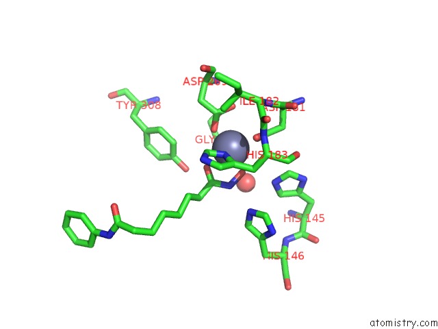
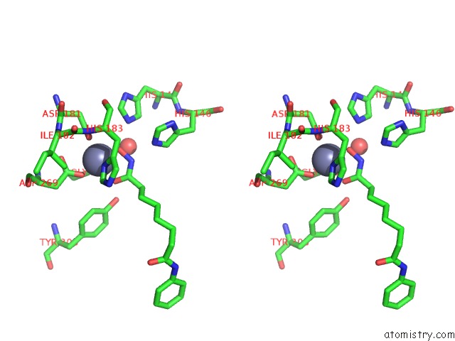
Zinc binding site 1 out of 3 in 4lxz
Go back to
Zinc binding site 1 out
of 3 in the Structure of Human HDAC2 in Complex with Saha (Vorinostat)
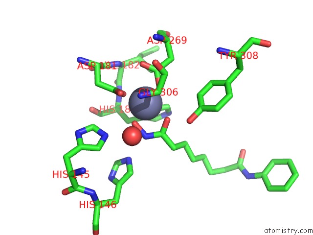
Mono view
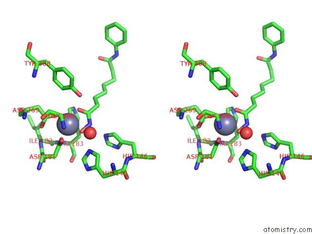
Stereo pair view
Mono view
Stereo pair view
A full contact list of Zinc with other atoms in the Zn binding
site number 1 of Structure of Human HDAC2 in Complex with Saha (Vorinostat) within 5.0Å range:
|
Zinc binding site 2 out of 3 in 4lxz
Go back to
Zinc binding site 2 out
of 3 in the Structure of Human HDAC2 in Complex with Saha (Vorinostat)
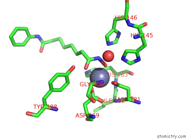
Mono view
Stereo pair view
Mono view
Stereo pair view
A full contact list of Zinc with other atoms in the Zn binding
site number 2 of Structure of Human HDAC2 in Complex with Saha (Vorinostat) within 5.0Å range:
|
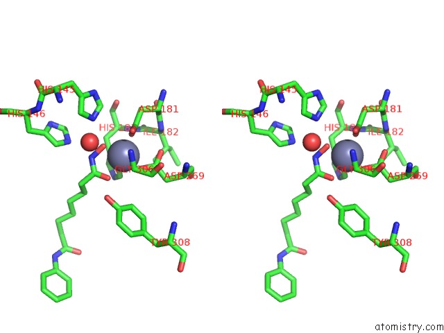
Zinc binding site 3 out of 3 in 4lxz
Go back to
Zinc binding site 3 out
of 3 in the Structure of Human HDAC2 in Complex with Saha (Vorinostat)
Mono view
Stereo pair view
Mono view
Stereo pair view
A full contact list of Zinc with other atoms in the Zn binding
site number 3 of Structure of Human HDAC2 in Complex with Saha (Vorinostat) within 5.0Å range:
|
Reference:
B.E.Lauffer,
R.Mintzer,
R.Fong,
S.Mukund,
C.Tam,
I.Zilberleyb,
B.Flicke,
A.Ritscher,
G.Fedorowicz,
R.Vallero,
D.F.Ortwine,
J.Gunzner,
Z.Modrusan,
L.Neumann,
C.M.Koth,
P.J.Lupardus,
J.S.Kaminker,
C.E.Heise,
P.Steiner.
Histone Deacetylase (Hdac) Inhibitor Kinetic Rate Constants Correlate with Cellular Histone Acetylation But Not Transcription and Cell Viability. J.Biol.Chem. V. 288 26926 2013.
ISSN: ISSN 0021-9258
PubMed: 23897821
DOI: 10.1074/JBC.M113.490706
Page generated: Sun Oct 27 02:08:02 2024
ISSN: ISSN 0021-9258
PubMed: 23897821
DOI: 10.1074/JBC.M113.490706
Last articles
Mg in 6VRFMg in 6VS4
Mg in 6VR8
Mg in 6VR7
Mg in 6VQP
Mg in 6VQT
Mg in 6VQA
Mg in 6VQ9
Mg in 6VQ7
Mg in 6VQ6