Zinc »
PDB 4gyf-4h8q »
4h01 »
Zinc in PDB 4h01: The Crystal Structure of Di-Zn Dihydropyrimidinase From Tetraodon Nigroviridis
Protein crystallography data
The structure of The Crystal Structure of Di-Zn Dihydropyrimidinase From Tetraodon Nigroviridis, PDB code: 4h01
was solved by
Y.C.Hsieh,
M.C.Chen,
C.C.Hsu,
S.I.Chan,
Y.S.Yang,
C.J.Chen,
with X-Ray Crystallography technique. A brief refinement statistics is given in the table below:
| Resolution Low / High (Å) | 30.00 / 2.00 |
| Space group | I 41 2 2 |
| Cell size a, b, c (Å), α, β, γ (°) | 160.478, 160.478, 93.296, 90.00, 90.00, 90.00 |
| R / Rfree (%) | 17.6 / 21.1 |
Zinc Binding Sites:
The binding sites of Zinc atom in the The Crystal Structure of Di-Zn Dihydropyrimidinase From Tetraodon Nigroviridis
(pdb code 4h01). This binding sites where shown within
5.0 Angstroms radius around Zinc atom.
In total 2 binding sites of Zinc where determined in the The Crystal Structure of Di-Zn Dihydropyrimidinase From Tetraodon Nigroviridis, PDB code: 4h01:
Jump to Zinc binding site number: 1; 2;
In total 2 binding sites of Zinc where determined in the The Crystal Structure of Di-Zn Dihydropyrimidinase From Tetraodon Nigroviridis, PDB code: 4h01:
Jump to Zinc binding site number: 1; 2;
Zinc binding site 1 out of 2 in 4h01
Go back to
Zinc binding site 1 out
of 2 in the The Crystal Structure of Di-Zn Dihydropyrimidinase From Tetraodon Nigroviridis
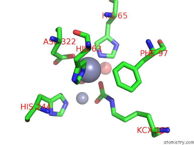
Mono view
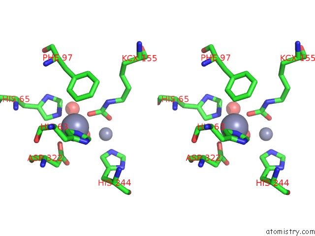
Stereo pair view
Mono view
Stereo pair view
A full contact list of Zinc with other atoms in the Zn binding
site number 1 of The Crystal Structure of Di-Zn Dihydropyrimidinase From Tetraodon Nigroviridis within 5.0Å range:
|
Zinc binding site 2 out of 2 in 4h01
Go back to
Zinc binding site 2 out
of 2 in the The Crystal Structure of Di-Zn Dihydropyrimidinase From Tetraodon Nigroviridis
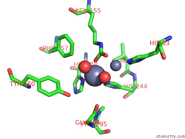
Mono view
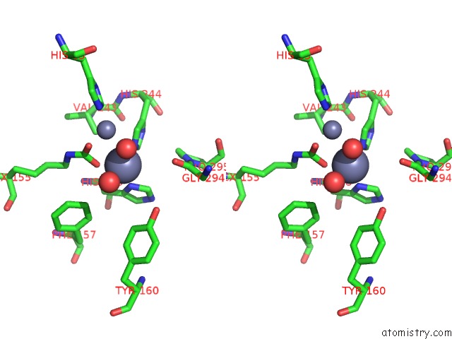
Stereo pair view
Mono view
Stereo pair view
A full contact list of Zinc with other atoms in the Zn binding
site number 2 of The Crystal Structure of Di-Zn Dihydropyrimidinase From Tetraodon Nigroviridis within 5.0Å range:
|
Reference:
Y.C.Hsieh,
M.C.Chen,
C.C.Hsu,
S.I.Chan,
Y.S.Yang,
C.J.Chen.
Lysine Carboxylation: Metal and Structural Requirements For Post-Translational Modification To Be Published.
Page generated: Sat Oct 26 23:45:14 2024
Last articles
Mg in 6CAPMg in 6CA4
Mg in 6C90
Mg in 6CA0
Mg in 6C9Y
Mg in 6C8Z
Mg in 6C8P
Mg in 6C8N
Mg in 6C8O
Mg in 6C8D