Zinc »
PDB 4cj0-4csz »
4ck0 »
Zinc in PDB 4ck0: Crystal Structure of the Integral Membrane Diacylglycerol Kinase - Form 2
Enzymatic activity of Crystal Structure of the Integral Membrane Diacylglycerol Kinase - Form 2
All present enzymatic activity of Crystal Structure of the Integral Membrane Diacylglycerol Kinase - Form 2:
2.7.1.107;
2.7.1.107;
Protein crystallography data
The structure of Crystal Structure of the Integral Membrane Diacylglycerol Kinase - Form 2, PDB code: 4ck0
was solved by
D.Li,
J.A.Lyons,
L.Vogeley,
D.Aragao,
M.Caffrey,
with X-Ray Crystallography technique. A brief refinement statistics is given in the table below:
| Resolution Low / High (Å) | 53.006 / 2.92 |
| Space group | P 31 2 1 |
| Cell size a, b, c (Å), α, β, γ (°) | 72.820, 72.820, 195.670, 90.00, 90.00, 120.00 |
| R / Rfree (%) | 24.79 / 28.09 |
Zinc Binding Sites:
The binding sites of Zinc atom in the Crystal Structure of the Integral Membrane Diacylglycerol Kinase - Form 2
(pdb code 4ck0). This binding sites where shown within
5.0 Angstroms radius around Zinc atom.
In total 2 binding sites of Zinc where determined in the Crystal Structure of the Integral Membrane Diacylglycerol Kinase - Form 2, PDB code: 4ck0:
Jump to Zinc binding site number: 1; 2;
In total 2 binding sites of Zinc where determined in the Crystal Structure of the Integral Membrane Diacylglycerol Kinase - Form 2, PDB code: 4ck0:
Jump to Zinc binding site number: 1; 2;
Zinc binding site 1 out of 2 in 4ck0
Go back to
Zinc binding site 1 out
of 2 in the Crystal Structure of the Integral Membrane Diacylglycerol Kinase - Form 2
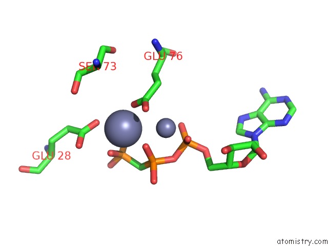
Mono view
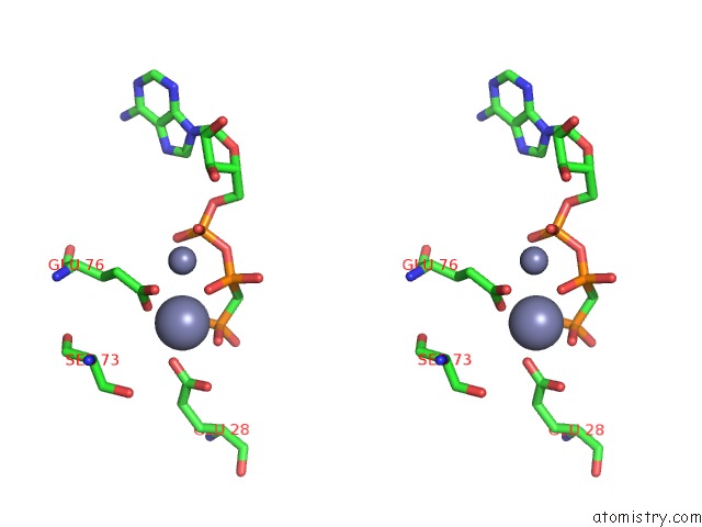
Stereo pair view
Mono view
Stereo pair view
A full contact list of Zinc with other atoms in the Zn binding
site number 1 of Crystal Structure of the Integral Membrane Diacylglycerol Kinase - Form 2 within 5.0Å range:
|
Zinc binding site 2 out of 2 in 4ck0
Go back to
Zinc binding site 2 out
of 2 in the Crystal Structure of the Integral Membrane Diacylglycerol Kinase - Form 2
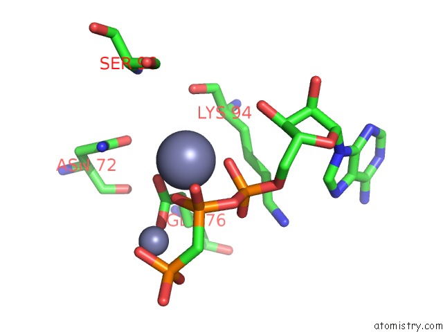
Mono view
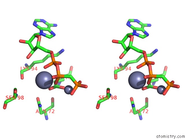
Stereo pair view
Mono view
Stereo pair view
A full contact list of Zinc with other atoms in the Zn binding
site number 2 of Crystal Structure of the Integral Membrane Diacylglycerol Kinase - Form 2 within 5.0Å range:
|
Reference:
D.Li,
A.Others,
M.Caffrey.
Crystal Structure of the Integral Membrane Diacylglycerol Kinase with Zn-Amppcp Bound and Its Catalytic Mechanism To Be Published.
Page generated: Wed Aug 20 16:45:57 2025
Last articles
Zn in 4K90Zn in 4K8B
Zn in 4K7W
Zn in 4K89
Zn in 4K88
Zn in 4K87
Zn in 4K7U
Zn in 4K86
Zn in 4K6T
Zn in 4K7S