Zinc »
PDB 4bjb-4bud »
4bm9 »
Zinc in PDB 4bm9: Structure of the Autoinhibited Parkin Catalytic Domain
Protein crystallography data
The structure of Structure of the Autoinhibited Parkin Catalytic Domain, PDB code: 4bm9
was solved by
T.Wauer,
D.Komander,
with X-Ray Crystallography technique. A brief refinement statistics is given in the table below:
| Resolution Low / High (Å) | 48.619 / 2.25 |
| Space group | H 3 2 |
| Cell size a, b, c (Å), α, β, γ (°) | 168.420, 168.420, 97.181, 90.00, 90.00, 120.00 |
| R / Rfree (%) | 19.03 / 21.78 |
Zinc Binding Sites:
The binding sites of Zinc atom in the Structure of the Autoinhibited Parkin Catalytic Domain
(pdb code 4bm9). This binding sites where shown within
5.0 Angstroms radius around Zinc atom.
In total 8 binding sites of Zinc where determined in the Structure of the Autoinhibited Parkin Catalytic Domain, PDB code: 4bm9:
Jump to Zinc binding site number: 1; 2; 3; 4; 5; 6; 7; 8;
In total 8 binding sites of Zinc where determined in the Structure of the Autoinhibited Parkin Catalytic Domain, PDB code: 4bm9:
Jump to Zinc binding site number: 1; 2; 3; 4; 5; 6; 7; 8;
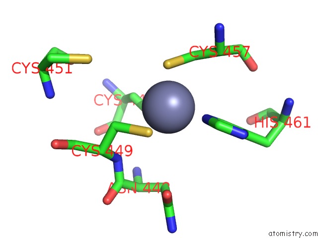
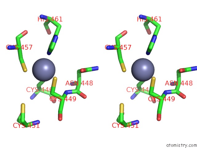
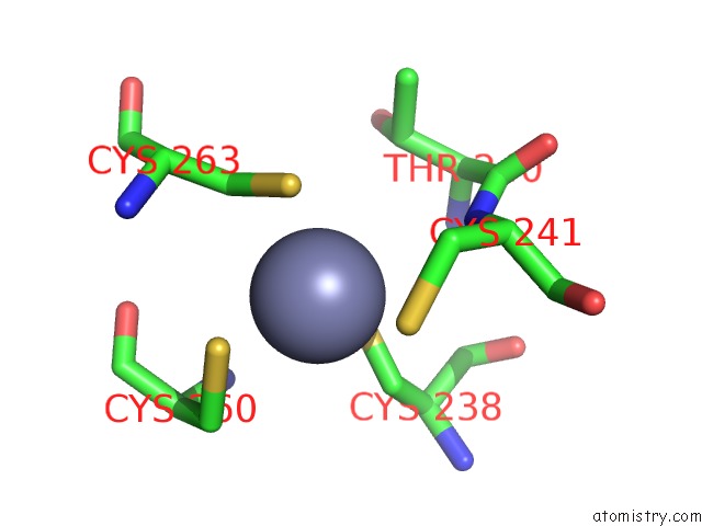
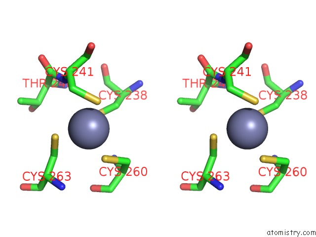
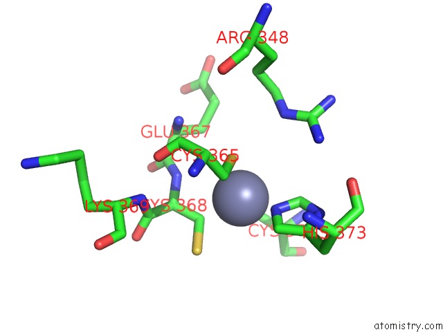
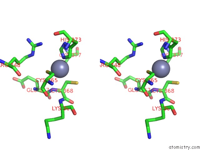
Zinc binding site 1 out of 8 in 4bm9
Go back to
Zinc binding site 1 out
of 8 in the Structure of the Autoinhibited Parkin Catalytic Domain
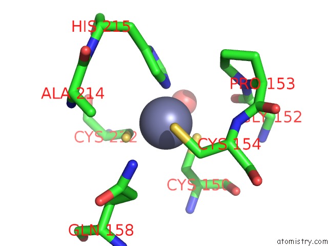
Mono view
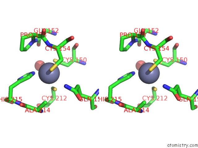
Stereo pair view
Mono view
Stereo pair view
A full contact list of Zinc with other atoms in the Zn binding
site number 1 of Structure of the Autoinhibited Parkin Catalytic Domain within 5.0Å range:
|
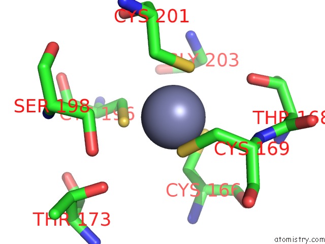
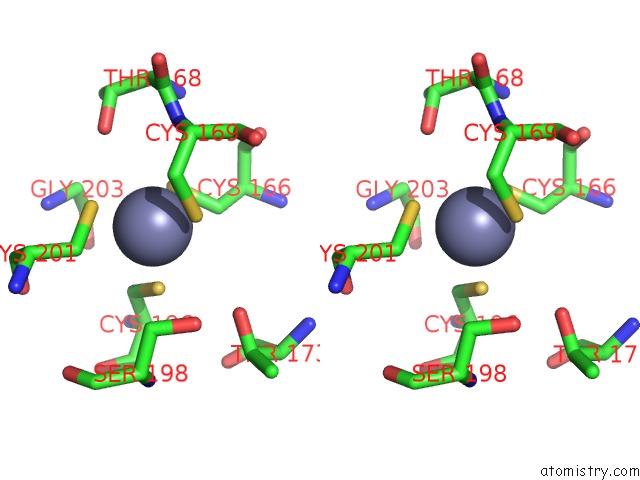
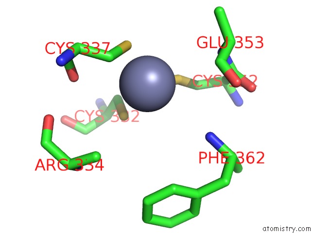
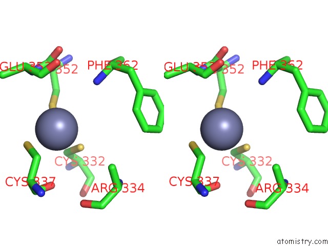
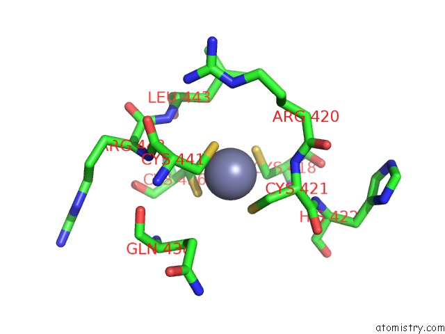
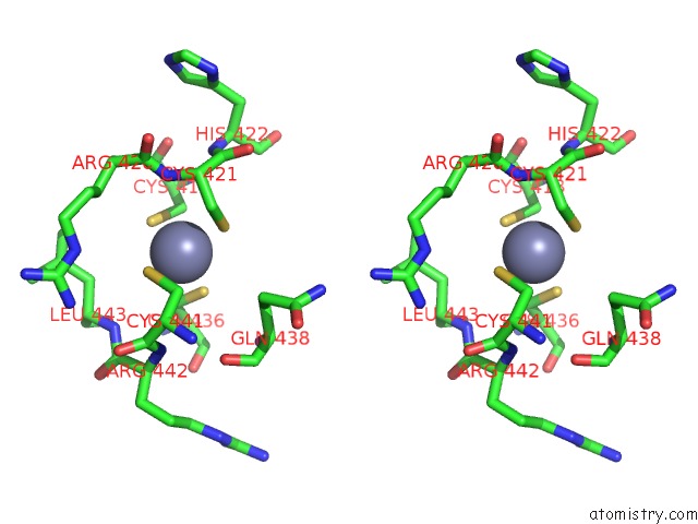
Zinc binding site 2 out of 8 in 4bm9
Go back to
Zinc binding site 2 out
of 8 in the Structure of the Autoinhibited Parkin Catalytic Domain
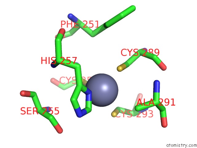
Mono view
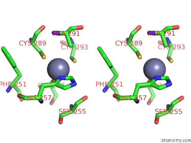
Stereo pair view
Mono view
Stereo pair view
A full contact list of Zinc with other atoms in the Zn binding
site number 2 of Structure of the Autoinhibited Parkin Catalytic Domain within 5.0Å range:
|
Zinc binding site 3 out of 8 in 4bm9
Go back to
Zinc binding site 3 out
of 8 in the Structure of the Autoinhibited Parkin Catalytic Domain
Mono view
Stereo pair view
Mono view
Stereo pair view
A full contact list of Zinc with other atoms in the Zn binding
site number 3 of Structure of the Autoinhibited Parkin Catalytic Domain within 5.0Å range:
|
Zinc binding site 4 out of 8 in 4bm9
Go back to
Zinc binding site 4 out
of 8 in the Structure of the Autoinhibited Parkin Catalytic Domain
Mono view
Stereo pair view
Mono view
Stereo pair view
A full contact list of Zinc with other atoms in the Zn binding
site number 4 of Structure of the Autoinhibited Parkin Catalytic Domain within 5.0Å range:
|
Zinc binding site 5 out of 8 in 4bm9
Go back to
Zinc binding site 5 out
of 8 in the Structure of the Autoinhibited Parkin Catalytic Domain
Mono view
Stereo pair view
Mono view
Stereo pair view
A full contact list of Zinc with other atoms in the Zn binding
site number 5 of Structure of the Autoinhibited Parkin Catalytic Domain within 5.0Å range:
|
Zinc binding site 6 out of 8 in 4bm9
Go back to
Zinc binding site 6 out
of 8 in the Structure of the Autoinhibited Parkin Catalytic Domain
Mono view
Stereo pair view
Mono view
Stereo pair view
A full contact list of Zinc with other atoms in the Zn binding
site number 6 of Structure of the Autoinhibited Parkin Catalytic Domain within 5.0Å range:
|
Zinc binding site 7 out of 8 in 4bm9
Go back to
Zinc binding site 7 out
of 8 in the Structure of the Autoinhibited Parkin Catalytic Domain
Mono view
Stereo pair view
Mono view
Stereo pair view
A full contact list of Zinc with other atoms in the Zn binding
site number 7 of Structure of the Autoinhibited Parkin Catalytic Domain within 5.0Å range:
|
Zinc binding site 8 out of 8 in 4bm9
Go back to
Zinc binding site 8 out
of 8 in the Structure of the Autoinhibited Parkin Catalytic Domain
Mono view
Stereo pair view
Mono view
Stereo pair view
A full contact list of Zinc with other atoms in the Zn binding
site number 8 of Structure of the Autoinhibited Parkin Catalytic Domain within 5.0Å range:
|
Reference:
T.Wauer,
D.Komander.
Structure of the Human Parkin Ligase Domain in An Autoinhibited State. Embo J. V. 32 2099 2013.
ISSN: ISSN 0261-4189
PubMed: 23727886
DOI: 10.1038/EMBOJ.2013.125
Page generated: Sat Oct 26 19:50:36 2024
ISSN: ISSN 0261-4189
PubMed: 23727886
DOI: 10.1038/EMBOJ.2013.125
Last articles
Mg in 4JI1Mg in 4JI0
Mg in 4JI2
Mg in 4JI3
Mg in 4JHD
Mg in 4JH6
Mg in 4JH8
Mg in 4JH7
Mg in 4JH3
Mg in 4JH5